Hemangiomas in children can be absorbed themselves or demand immediate medical intervention. How the type and treatment of hemangiomas in children is determined, this article will tell.
Content
- Hemangioma in children. The reasons
- Hemangioma in children photo
- Hemangioma on the head of a child
- Hemangioma on the lip in a child
- Hemangioma on the face of a child
- Hemangioma on the back of a child
- Hemangioma in children up to a year
- Hemangioma in newborns
- Skin hemangioma in children
- Subcutaneous hemangioma in a child
- Vascular hemangioma in children
- Liver hemangioma in children
- Cavernous hemangioma in children
- Hemangiom treatment in children
- Removal of hemangioma with a laser in children
- Video: What is hemangioma - a school of Dr. Komarovsky?
One of the ailments of the infant period of the child’s life is hemangioma - a tumor -shaped vice of blood vessels, outwardly resembling a dirty stain on the skin.

Such “spots” can have different colors: from pale pink to crimson, but most often there are hemangiomas of reddish-blue shades.
Important: hemangiomas may not appear on the child’s body immediately, but only after 1 - 2 months from birth. In female infants, hemangiomas occur slightly more often than in babies-boys.

Hemangioma in children. The reasons
The exact causes of the formation and growth of hemangiomas are not known to this day, but with a high probability they are:
- colds and viral diseases of a woman at an early gestation (up to 12 - 14 weeks)
- adverse environmental living conditions for the future mother
- multiple pregnancy
- strong non -prematureness of the child
- mom's age is older than 32 - 35 years old (the older a pregnant woman, the higher the risk of hemangiomas in the fetus)
- fetoplacental failure
Important: there is an opinion that the way of gemangias in infants is affected by the method of childbirth. According to statistics, children born by cesarean section are observed more often than in babies born naturally.

Hemangioma in children photo
Hemangiomas in children can be small and bright. They, as a rule, rarely cause inconvenience and almost never grow.

However, there are such hemangiomas, the appearance and location of which causes a lot of problems and suffering in the child.

Hemangioma on the head of a child
Hemangioma on the head of a child is a very frequent phenomenon. This benign tumor can appear anywhere in the skull. Hemangiomas located on the head are dangerous to close neighborhood with the brain, eyes, ears and respiratory organs.
Important: the first signs of the formation of hemangioma on the head may be the swelling of the skin and a slight change in its color.
Gemangiomas of the heads require medical control. If the tumor, increasing, begins to squeeze vital organs, the doctor must decide to remove it.
Hemangioma on the lip in a child
In addition to the unaesthetic species, the hemangioma located on the lips can cause the child inconvenience when taking and chewing food. Babies born with the hemangiomas of the lips often refuse breastfeeding, as they cannot correctly capture the nipple.
Important: if hemangioma has growth trends, it can eventually go beyond the lips and “spread” to the chin, cheeks or nasolabial folds.
Two methods can be used to remove hemangiomas from the child's lips:
- restless laser operation (for capillary hemangiomas)
- burning with liquid nitrogen (for cavernous and mixed forms of hemangioma)

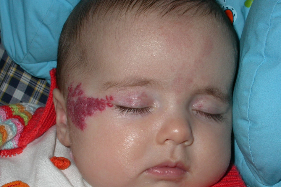
Hemangioma on the face of a child
Hemangioma on the face of a child is sometimes a danger to the normal functioning of the organs of vision, smell and hearing.
In addition, it is a serious cosmetic defect that over time can make the child feel "not like everyone else." Regardless of what type, color and form has a hemangioma on the face, it will certainly attract the sympathetic views of others.
Important: in cases of minor external manifestations of the defect, doctors recommend that parents not rush with the operation, but watch the condition of the affected area of \u200b\u200bthe skin for some time. Often hemangiomas on the face are absorbed themselves, without any medical intervention.

Hemangioma on the back of a child
Hemangiomas on the back appear in 19 children out of 100. Most of them are congenital. They are formed on the child’s body when he is still in the mother womb and can be located on the lower back, spine, ribs or shoulder blades.
In most cases, hemangiomas on the back are not dangerous for the health and life of children. Capillary formations can grow, or can, on the contrary, decrease and pale. Cavernous and mixed hemangiomas do not change their appearance and sizes and often germinate into the subcutaneous layers.
Important: hemangiomas on the back do not degenerate into malignant tumors, so with surgical removal they can be held. By the 5th - 7th year of age, the back hemangiomas independently disappear without a trace in many children.

Hemangioma in children up to a year
When hemangiomas are found in children up to a year, parents often panic. In fact, everything is not as scary as it might seem right away.
If the dimensions, form and arrangement of hemangioma do not cause suspicion among doctors, parents just need to observe it and mark any changes.
Important: 2% of children are born with hemangiomas, and in 10% of babies they are formed during the first year of life. Of all cases, 95% are simple (capillary) hemangiomas.

Hemangioma in newborns
Hemangiomas in newborns are a frequent phenomenon. The main reason for the appearance of hemangiomas on the body of the fetus located in the womb is a violation of the normal formation of the vascular system (3-6 weeks of pregnancy).
Typically, the size of hemangiomas in newborns does not exceed 2 cm, but there are exceptions when skin lesions are extensive and numerous. In most cases, all of them are absorbed themselves during the first years of the child’s life.
Important: treatment with hemangiomas in newborns primarily implies the observation of the child with a pediatrician, dermatologist and surgeon.

Skin hemangioma in children
Hemangioma of the skin in children there are 2 species:
- congenital - if the child was born with hemangioma
- nursery - if the defect appeared some time after birth
Congenital hemangiomas usually do not change in size and resolve themselves until the age of ten. Children's hemangiomas can either increase or decrease until they disappear without a trace.
Important: the diagnosis of hemangioma cannot be made independently. If you notice an unusual reddish spot on the child’s skin, contact a dermatologist to clarify the nature of the defect.
The presence of a child on the skin of a child in most cases does not affect his physical and mental development.

Subcutaneous hemangioma in a child
The subcutaneous hemangioma has clear boundaries on the surface of the skin and differs in red or blue. A bright stain on the skin will turn pale if you press a little finger on it.
This happens due to a quick outflow of blood. Subcutaneous hemangioma to the touch is slightly warmer than healthy skin.
Important: subcutaneous hemangioma is dangerous to the likelihood of complications in the form of bleeding, phlebitis and thrombophlebitis in case of damage.

Vascular hemangioma in children
Vascular hemangioma consists of blood vessels, lymph nodes and other tissues. Outwardly, it manifests itself on the skin of a child, like red or blue spots, from 0.5 to 10 cm in size. Most often localized in the head.
The color and consistency of formations are determined by fabrics that are part of the tumor. On the skin area with vascular hemangioma, blood supply is impaired, but this should not scare the parents of the child, since such defects are prone to self -removal.

Liver hemangioma in children
The hemangioma of the liver in a child may not be seen by parents. If its dimensions do not exceed 5 cm, then no symptoms of its presence can be detected. Most likely, over time, she will resolve and will never remind herself again.
If the dimensions of the hemangioma of the liver over time increased to 10 cm or more, the child will complain about aching pains in the right hypochondrium, a feeling of composure in the stomach and intestines.
Important: from the benign tumor of the hemangioma of the liver, which is a ball of blood vessels, can be transformed into malignant. The rupture of the hemangioma of the liver is also dangerous-because of this, the child may have internal bleeding.
If hemangioma of the liver is detected in a child, parents should exclude spicy, fatty, smoked, salted and fried dishes from his diet. In return, you can offer strawberries, carrots, beets, fish, liver or dairy products.

Cavernous hemangioma in children
Cavernous hemangioma - a formation consisting of two or more vascular cavities filled with blood. It grows from the subcutaneous fat layer. If the cavernous hemangioma begins to grow, the skin covering its skin will acquire a bluish-gag tint.
Only a doctor can diagnose a hemangioma of this species based on the results of ultrasound and laboratory tests.
Important: if a child was diagnosed with cavernous hemangioma, it should be immediately treated to its treatment.

Hemangiom treatment in children
According to statistics, in 10% of children, hemangiomas are absorbed by the year, in 50% - to five, and in 70% - by 7 years. However, if hemangioma increases in size or threatens the child’s health, it is necessary to engage in treatment.
Hemangiomas can be treated with one of the following methods:
- with the help of medicines
- cryotherapy
- casualties
- injections of sclerosing substances
- laser therapy
- with the help of folk remedies
Important: in each individual case, treatment is selected individually depending on the type, size and degree of severity of the defect.

Removal of hemangioma with a laser in children
Modern removal of hemangiomas in children is a painless effect on the affected area of \u200b\u200bthe skin with a laser.
The laser “reduces” the tumor to the minimum size, while eliminating traces from it. After this procedure, wounds heal very quickly, without complications.
IMPORTANT: Removing by hemangiomas with a laser is the safest way to solve the problem. The result of the removal procedure depends only on the qualifications of the surgeon conducting it.
The disadvantage of this method is the inability to remove the hemangioma at a time. On average, for the complete disappearance of the defect, 3-15 procedures with an interval of 2 to 3 weeks will be needed.

If you notice a suspicious spot on your child’s body, according to the description similar to a hemangioma, do not despair. Show the child to an experienced doctor who, upon examination, will accurately determine the origin of the skin defect and, if necessary, prescribe adequate treatment.