Arthrosis affects the joint and interferes with the normal movement. Let's figure out how to get rid of an unpleasant ailment.
Content
- How the change in the joints is expressed: the first signs of arthrosis of the hip joint
- Symptoms with deforming arthrosis of the hip joint
- Causes causing arthrosis of the hip joint
- How to diagnose arthrosis of the hip joint?
- What is treated by arthrosis of the hip joint?
- Signs of different degrees of arthrosis of the hip joint
- Therapeutic gymnastics for arthrosis of the hip joint: indicated for the use of patients of the first and second group
- Video: Coksartrosis: Reason, treatment. Arthrosis of the hip joint - symptoms of 1, 2 or 3 stages
Arthrosis is a systemic disease of the joints. It is divided into deforming, basic and congenital (genuine). This is perhaps a predominantly common disease of articular bags. The basis of this disease is aging and a change in joint cartilage, associated with a decrease in the cartilage of its most important component - proteoglycan.
How the change in the joints is expressed: the first signs of arthrosis of the hip joint
- The degeneration and change in cartilage is caused by the seal and deformation of the joints of the joints with some areas of deadlife in it (cyst), as well as a huge growth of the edges of the joints, to which changes in the synovial shell, sclerosis and a decrease in joint capsule are attached. All these changes lead to constant deformation of the joints, but without a sharp violation of their functions.
- The joint is a living structure; Its functions depend on the content of various substances and the state of the joint capsule. Arthrosis, just changes this very, articular capsule and, in general, the entire joint.
- Basically, the joints of the pelvis and hips (hip), also joints of the knees, joints between the flanks of the hands, but arthrosis is primary, are the first to hit. With damage to the joints of the pelvis and hips, it is also called - coxarthrosis or deforming arthrosis.

- Everything begins with the appearance of unstable pain in the joints in the evening. As well as slowly occurring on the increasing deformation of the joints due to bone growths.
- In case of insufficient inflammatory process, a small, barely pronounced pain and unstable swelling (reactive synovitis) occurs occurs in the articular fabric (swelling).
- When a dead piece of bone tissue breaks off, and it is infringed between the surfaces inside the joint, the so -called “blockade symptom” can occur - terribly acute joint pain with intensity in the joints, and it passes sharply quite sharply. The total body temperature and blood indicators will be completely normal.
Symptoms with deforming arthrosis of the hip joint
- The first mainly, some growing pain is manifesting closer to evening time of the day.
- The appearance of bursting pain. As it were, pain occurs from the inside and moves to the surface.
- The surface swells around the joint, often this edema passes by itself.

Pain and swelling - There is a sharp pain in the joint, and passes quickly.
- It is completely impossible to move the joint.
- The area around the joint is distorted, which becomes visible with a simple look.

Causes causing arthrosis of the hip joint
- The professions associated with the load on this joint are movers, ballerinas, foot couriers, professional athletes.
- Overloading the joints with older people. This type of overload is quite common. Pensioners living alone should take care of pressing problems. And this leads to the fact that they overload their joints.
Overload, injuries, age - main reasons - Overload of the joints after, any inflammation. Perhaps the person suffered arthritis and then loads the joint. This cannot be done categorically.
- Injuries and microtrauma of the joints. This reason is quite common, 35% of patients with arthrosis suffered any injury. And after that, something incomprehensible happens with the hip joint.

How to diagnose arthrosis of the hip joint?
IMPORTANT: At the first unpleasant sensations in the area of \u200b\u200bthe hip joints, contact the therapist.
- If you hear a characteristic crunch in the joint, pay attention to this. The crunch is deaf, not very sonorous.
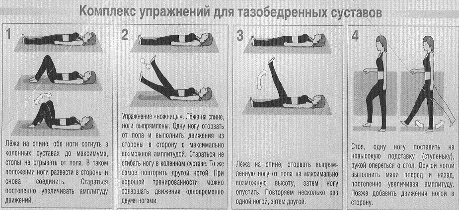
- Lie on the floor, raise your legs up and make a circular movement with your foot to the side, down, and then up. If, when you do this exercise, you feel that the joint comes out of its place, and then returns with a click, consult a doctor. Tell us about it.
- You can do the same exercise standing: bend your leg at the knee and lift it to the pelvis, scroll your foot forward and backward. Follow your feelings.
- Also lie on the floor and do the exercise "scissors". At the same time, the joints should not hurt.
- It is best to make an X -ray to make a x -ray in case of suspicion of arthrosis of the hip joint. The picture will best tell about the changes that are taking place.

- You can also make an MRI, in which case you will get a full picture in different projections of your joint.
- There is also a special device that shows the mobility of the joint (goniometer). With it, you can diagnose joint mobility.
- Drive the blood for analysis, you need to do a detailed blood test in order for the doctor to pay attention to the intended changes.
- You also need to hand over the urine analysis. The high content of proteins in the urine speaks of the inflammatory process in the body.
What is treated by arthrosis of the hip joint?
It is necessary to treat arthrosis of the hip joint as soon as it manifested itself, and it is better to prevent it. How preventive agents use chondroprotectors. They are prescribed to drink, and professional athletes, despite their age, and just older people to maintain cartilage fabric in normal.
Chondroprotectors include:
- chondroitin
- chondroitin with glucosamine
- teraflex Advance
- alflutop
- structure
- rumalon
- don and so on
But when you are diagnosed: “coksoarthrosis of the hip joint”, then you need shock therapy. Tablets, an analgesic (NSAID), and injections are used.

The use of simple analgesic agents, with an anti -inflammatory effect, relieves pain in most cases. In severe inflammation, injections of more powerful anti -inflammatory drugs are prescribed, directly into the affected joint. Or surgically, replacing the joint with artificial.
-
Analgesics
Analgesics are drugs to relieve pain. Damage to the tissue for a disease or injury is perceived by nerve endings that transmit signals to the brain.
Analgesics are divided into opioid and neopioid. Neopioid drugs include all other analgesics: paracetamol, non -ifopam, non -steroidal anti -inflammatory drugs, from which aspirin is most known. Neopioid analgesics are less active. Local anesthetics are also used to eliminate pain.
-
Non -steroidal anti -inflammatory drugs (NSAIDs)
NSAID Aspirin. For many decades have been used to relieve pain and fever. It also reduces inflammation, helps to reduce swelling and pain. Aspirin in combination with other drugs is part of many drugs. However, it affects blood coagulation and therefore does not suit patients with poor blood coagulation. For the treatment of articular pain, diclofenac, diclobert capsule, ibuprofen, indomethacin, ketoprofen, meloxicam are used.
NSAIDs Metaminic acid. The drug presented in 1963. Relieves pain, prescribed with inactivity of the joints. The most common side effects are observed from the gastrointestinal tract: abdominal pain, nausea, vomiting and indigestion.
-
Rheumatic pain preparations
They are used to treat rheumatic diseases. In the disease, pain, stiffness and swelling of the joints are noted, and with a long course, their deformation is possible. The treatment is aimed at relieved symptoms of pain and stiffness in the joints, maintaining mobility and preventing joint deformation.

Indomethacin, a budodion of 0.025 g 3-6 times a day, deeds 0.25 g times a day, preferably at night. After one and a half to two months, breaks should be taken in treatment for 10-15 days. There may be intra -articular administration of hydrocortisone. Azatioprine, cyclosporine, cyclophosphamide, leflunomide, methotrexate, and vlokortolone are also used.
-
Corticosteroda for local introduction
The adrenal glands produce a number of important hormones. Among them are corticosteroids, named so because they are produced by the outer, cortical part of these glands. Corticosteroids affect the immune system and regulate carbohydrate and mineral metabolism in the body. A number of drugs imitating natural corticosteroids have been developed. This article contains information about corticosteroids used for injection in the affected areas for the treatment of joint diseases.
Local injections of corticosteroids are used to reduce pain and inflammation. Sometimes they can be prescribed to reduce the severity of symptoms sufficient for physiotherapy. These include: prenisolone, triamcinone, dexamethasone, hydrocortisone, methylprednisolone.
-
Expanding vessels drugs
With swelling or change in the joints, the vessels that surround them are squeezed, and provide the damaged joint with nutrition. This side of the issue is resolved by the use of special vasodilating drugs. Such as: Actovegin, Penctosiphilin, Eufillin and Lipolic acid, in some cases, no-shpu 2 tablets are prescribed three times a day. The vessels expand, and the blood system can actively nourish the sore surface, this greatly helps in the treatment of arthrosis of the hip joints.

It is interesting that recently a method has become popular in which the blood (plasma of blood) of the patient, who was enriched with platelets, is introduced into the articular fluid. Due to the fact that the “medicine” is produced by the patient himself by no side effects. The method is called - plasmolifting.
Signs of different degrees of arthrosis of the hip joint
The three degrees of arthrosis of the hip joints are recognized by doctors:
- First degree: The pain is not very large, moderate, joint mobility is not limited. On the X -ray shows a slight narrowing of the joint bag, there is no growth of joint tissue, osteophytes can be slightly visible. If you start treatment in time, the first degree is perfectly corrected by specialists. The patient can be cured.
- Second degree: There is constant pain in the joint, even at rest. A person cannot raise his leg and turn it aside. It is often accompanied by acute pain when turning the foot to the right and left. The X -ray examination shows a significant growth of the joint, numerous outgrowths, narrowing of the joint slit literally half. Timely treatment can suspend the course of the disease for a while.
- Third degree: The pain is constant, under no circumstances, atrophic changes in the bones and buttocks are visible, the patient is “skewed” to the side, where the joint is most affected. In case of illness of both joints, there is a “gait of a duck”. In the pictures of the X -ray, complete deformation of the joint is visible, the joint gap is not visible (complete narrowing). Treatment does not bring noticeable results, it is necessary to replace the joint surgically.
3 degrees

Therapeutic gymnastics for arthrosis of the hip joint: indicated for the use of patients of the first and second group
- Become straight legs shoulder -width apart, your back is flat. Put your hands on the belt, raise your right hand up and left. We tilt to the left, two or three pull-ups and slow down for thirty seconds. We make the same movement with the left hand to the right. 4-5 inclinations of 6-8 approaches.
- The starting position is also as in the first exercise. Hand your hands up above your head. We tilt forward, stretch our hands, as if we want to pick up something in our hands. Freeze for thirty seconds. We make 4-5 times in 6-8 approaches.
- Stone hands on the waist, legs shoulder -width apart. They bent their leg at the knee and raised 90 degrees in relation to the body. We make circular movements forward, and then back. 10-15 movements of 8-10 approaches for each leg.
- They began exactly in front of the wall or took up the back of the chair. We make swings with your foot to the sides and back. 30 slices each foot 3 approaches.
- We took up the back of the chair. We stand sideways to him, knelt to the sides, legs below in the first position. Squats at the expense of one to eight. We make 6-8 squats up to 10 approaches.
- We sat on the floor. The body forms an angle of 90 degrees. Sit exactly on both muscles of the pelvis. Alternately move one half of the body forward, after it is also the second. You are “walking” with a basin on the floor. 30-40 moves forward and the same amount back. 3 approach.
Some exercises - Lie on your back with your arms spread to the sides of the legs bend in your knees and lift over the floor. We make the “twisting” of the body, put our legs in turn, then to the right, then to the left. In this case, the upper body does not move at all. 20-30 times in each direction, three approaches.
- Lying on your back, do the exercise "Bicycle". They raised their legs above the floor and bent, in turn, straighten one leg along one leg, the second is bent. It looks like a bicycle ride. 20-30 times in 4-6 approaches.
- Lying on the back, take the right leg to the side, bend and straighten up, bend again and then return the right leg to the left leg along the body. How to slip your foot to the side and down. Exercise do 10-30 times in 4 sets.
- We got up. The back is even, legs shoulder -width apart. They raised their right leg, hugged her with two hands and pull the leg to the chest. We make 20-30 times 3 approaches to each leg.