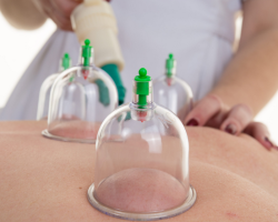
Now the study of the function of the cardiovascular and respiratory systems is carried out using radioactive isotopes. Read more in the article.
Content
- Heart and vascular diseases: list
- Study of the function of the human cardiovascular system using radioactive isotopes: Modern diagnosis of heart disease
- Study of the function of the lungs, the respiratory system of a person using radioactive isotopes
- Video: radiation diagnostics of the cardiovascular system (LD CCC)
- Video: The most common diseases of the cardiovascular system
- Video: prevention, diagnosis and treatment of cardiovascular diseases
Over the past decades, radioactive isotopes in clinical diagnostic practice are increasingly used in connection with achievements in physics, chemistry and radiobiology, successes in the construction of new radiometric equipment and obtaining the results of new compounds.
Read in another article on our website on the topic: "What to do with a heart attack at home?". You will learn about the symptoms of this condition, as well as how to provide first aid, you will find prevention advice.
In this article you will read information about a new and modern study using radioactive isotopes. You will know what substance such a procedure is performed and whether it is harmful to the human body. Read further.
Heart and vascular diseases: list

Practical cardiology develops in two directions:
- Therapeutic cardiology
- Cardiac surgery
Therapeutic cardiology uses conservative methods (medications, sanatorium treatment) for the treatment of heart disease and blood vessels - a list:
- Bradycardia
- Tachycardia
- Arrhythmia
- Extrasystole
- Vegeto-vascular dystonia
- Atherosclerosis of the vessels
- Arterial hypertension
- Angina pectoris
- Myocardial infarction
- Cardiac ischemia
- Heart failure
- Myocarditis
- Pericarditis
- Endocarditis
Surgical cardiology promptly eliminates congenital and acquired heart defects and other damage to the heart and blood vessels, performs the prosthetics of valves and vessels of the heart.
Study of the function of the human cardiovascular system using radioactive isotopes: Modern diagnosis of heart disease

As you know, the practice of clinical radio -diagnostic research is increasingly included a scanning method that allows you to obtain an image of organs or tissues accumulating certain compounds laid by radioactive isotopes. Read more about the study of the function of the human cardiovascular system using radioactive isotopes:
- The image is obtained using the device - a scanner, the moving sensor of which sequentially records the radiation of the radioactive isotope above all areas of the studied area.
- The electronic system of the device allows the registration device that moves synchronously with the sensor, to write in the form of strokes, points, numbers the number of pulses coming from the sensor.
- According to the scanogram, one can judge the size, form, position and partially of the function of the body under study. In addition, the scanogram allows you to determine the presence of foci of increased or reduced radioactivity.
Valuable data were obtained when scanning the heart J131 albumin. In addition to previously conducted studies on the differential diagnosis of pericarditis and heart dilatation, modern diagnostics of localization of thrombitized and unpleasant aneurysm of the heart became possible.
- Scanning myocardium is possible after the introduction of water -soluble fatty acids into the body - J131.
- Although the obtained scanograms reflect quite accurately the contours of the myocardium, they do not give sufficient grounds for the differential diagnosis between the true acute myocardial infarction and the picture observed during the thinning of myocardial tissue or its replacement after the heart attack with a scar tissue.
- Nevertheless, in this case, the obtained scanograms can serve as a valuable addition to other methods of diagnosis of the disease.
When studying hemodynamics, the determination of coronary blood flow is of great importance, which, according to the previously proposed methodology, was determined after the intravenous administration of the isotope by the third peak on a descending heart curve. At the same time, it was believed that the third peak reflects the transition of blood to the system of coronary arteries of the heart. According to the section of the exponential curve lying under the third peak and heart curve, the value of coronary blood flow was determined.
It is worth knowing: However, the opinions of many scientists in assessing the magnitude of the third peak differ, and therefore such a technique has not received a wide clinical distribution.
It is possible that it is more reliable methodology of menathat allows you to determine the ratio of the inclination of the blood radioactivity curve in the heart and brain after intravenous administration J131-albumin. This reflects the state of coronary blood flow and is expressed in such relative indicators as a coronary index. On average it is equal 1,54+0,18 and significantly decreases with severe coronary insufficiency - less than 1.0.
Interesting data were obtained in the study of atherosclerosis and thrombosis using J131-fibrinogen and J131-fibrinolysin. It was found that:
- These compounds accumulate in the foci of thrombosis and in atherosclerotic deposits in larger quantities than in normal tissue.
- Experimental studies with an external score of radioactivity made it possible to determine the location of thrombosis, and also to establish that the radioactivity of blood for atherosclerosis is lower than normal.
These observations allow you to think about the possibility of using this test for the clinical diagnosis of atherosclerosis.
Study of the function of the lungs, the respiratory system of a person using radioactive isotopes

It is known that in the diagnosis of heart pathologies, the functions of the lungs are also carried out in parallel. Indeed, without the normal work of this paired organ, the heart will not function well. To study the function of the lungs, except for the previously used methods with radioactive gases xenon and crypton, new methods of studying perfusion and ventilation of the lungs are proposed. The study of the functions of the lungs and the entire respiratory system is now also actively carried out using radioactive isotopes.
For the study of blood circulation in this organ, they use macro Agricults J131-albuminobtained from the usual all the same J131-albumin, heating it for a short time at t 79-80 ° C.
- Particles of albumin macro -aggregates of albumin macro 10-50 mk The capillary-alveolar network of the body for a period sufficient to scan in their path.
- The thin, delicate flakes of the albumin macro -units quickly break up in the lungs.
- After decay, the microegates pass into the bloodstream and are absorbed by the reticulo-endothelial system of the liver and spleen.
- There are no complications when applying this technique.
- Scanograms give a distinct idea of \u200b\u200bthe blood -filling of the lungs and the obstacles arising on the path of pulmonary blood flow.
To study pulmonary ventilation and the condition of pulmonary air routes, the method of scanning the lungs after aerosol inhalation of various radioactive isotopes is used - J131-Benalrosis, radioactive colloidal AU198, J131-Hippuran and etc.
It is worth knowing: Spraying and the introduction of radioactive aerosols into light particles using an electric inagenter.
How it works:
- The optimal value of particles for spraying - from 5 to 15 mk.
- The half -life of the drug from the lungs - about 2 hours.
- The dose of irradiation of the body and lungs is small.
- With partial or complete obstruction of the bronchial tree, the particles of aerosols do not penetrate the affected area, and the scanogram notes a partial or complete absence of radioactivity in the affected area.
The most appropriate from a clinical point of view is the use of both research methods in various forms of pulmonary pathology. The results of such combined studies showed that with emphysema, tuberculosis, pneumonia, cystic fibrosis and some other lung diseases, foci or areas of absence and radioactivity of both injected and inhaled particles are detected on the scanogram. In other diseases, the relationship changes - areas of deficiency of injected particles with a relatively normal inhalation scanogram are usually characteristic of impaired blood supply - pulmonary embolism, pulmonary hypoplasia. The reverse picture is observed in violation of the ventilation of the lungs - bronchial asthma, obliterating bronchiolitis.