If you study the anatomy and structure of the human heart, then read the article. It has a lot of useful information.
Content
- Where a person has a heart: photo
- Functions, human heart: list
- What is the structure of the human heart - the heart in the context, right, left atrium, walls, muscles: describe briefly, anatomy (biology 8 grade), drawing
- Cameras in the human heart - structure, quantity: table
- Valves in the human heart - internal structure: signature scheme
- What structure have a human heart vessels: description
- Hystology of the heart: what does this human organ look like under a microscope, table
- Circles of blood circulation of the human heart - a large, small circle: where and where does the blood move through the vessels?
- Human heart cycle
- Human heart: types, magnitude
- Human heart structure: age -related features
- Video: heart anatomy
The human heart is asymmetrically in the chest, with a slight displacement to the left of the midline. This is a small, equal to a clenched fist, a muscle organ surrounded by a shell on the outside with a pericardium or a pericardial bag. The heart, in order to supply the internal organs of a person with nutrition, daily pumps about seven tons of blood. It, throughout the entire life cycle, produces on average 2.55 billion beats.
In another article on our website you can study human anatomy - the structure and location of internal organs. It has a lot of useful information for study.
The final formation of the muscle organ occurs in the tenth week during the period of intrauterine development of the fetus. After birth, it radically changes the principles of hemodynamics - circulatory systems: from the power of the mother, the mother is transition to pulmonary, independent nutrition. From this article you will learn where the human heart is, what a structure has and a lot of other interesting and useful information. Read further.
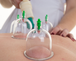
Where a person has a heart: photo

The main part of the human heart, or rather two -thirds of its volume, is on the left side of the center of the chest. And only one of the three parts enters the right half a little. This can be seen in the photo above. Read more:
- The heart in the human body is placed between his lungs.
- It closely adjacent from the inside to the chest and is surrounded by a corrugation bag.
- At the same time, the heart "axis" has a small slope.
- This location is considered the norm and is found in most people.
But deviations from the location of this body are possible:
- Right -sided location.
- Splasty in diaphragm or horizontal. The situation is possible with a wide, but short chest.
- The displacement is closer to the vertical location. It is found in thin people.
In addition, the position of the organ can curtain from the somatic constitution of a person. In the asthenic type of people, it is vertical. Hyperstenik has more often a horizontal location, the asthenic type is vertical.
Therefore, the location standard can undergo minor changes depending on the size of the heart, its individual characteristics, physiology of the human body and at the same time not to be considered a pathology. And therefore, it is possible only to talk about conditional standards, but not about the standard.
Functions, human heart: list

The heart is the main organ of the human body, and violations in its work are caused by further total disorders, and its stop leads to death. Several basic functions of the human heart are distinguished. Here is a list of functions and description of the work:
AUTOMATISM:
- This function is characterized by the independent development of electrical excitation pulses that contribute to the reduction of the heart muscle.
- Due to a violation of this connection, blood circulation and further death of a person are possible.
Conductivity:
- In the structure of the human heart are conductive paths that ensure the passage of the electric charge inside the heart muscle. But it does not act chaotic, but has a certain sequence - from the atrium to the ventricles.
- In case of violation of the relationship, pathological conditions may appear: the development of arrhythmias, heart rhythm disturbances, blockade.
- To correct the situation, medical therapeutic treatment will be required, and in extreme cases, surgical intervention.
REDUCTION:
- In most cases, the cells of the heart system provide muscular reduction.
- The mechanism of work is compared with the action of the muscles of the iris of the eye, biceps, and triceps - a signal of atypical cardiomyocytes enters the muscle, under its influence there is a reduction.
- In case of violations of muscle contractility, a person can observe various types of edema formed due to heart failure.
Pumping function:
- Provides full and timely pumping of blood under pressure through the vessels of the human body.
- Pressure is created in the process of reduction and relaxation of the heart.
- When malfunctions, the life of a person without additional medical care is impossible.
Oxygen power and supply:
- Blood enriched with nutrients and oxygen passes through the heart blood vessels with each heart contraction.
- The heart organ is a hollow muscle consisting of four chambers.
- The myocardial partition divides the heart into two parts, each of which has its own functionality. The right half passes venous blood, pushing it in the artery in the lungs. The left, receiving blood from the artery, enriched with nutrients and oxygen, takes it into aorta, then spreading through the body.
Endocrine function:
- In the right at the atrium, a sodium -reuters hormone is produced, which affects the functioning of the kidneys and the removal of harmful salts from the body.
- It stimulates the work of the kidneys, simultaneously contributing to a decrease in blood pressure.
Below is the structure of the human heart. Read further.
What is the structure of the human heart - the heart in the context, right, left atrium, walls, muscles: describe briefly, anatomy (biology 8 grade), drawing

The heart looks like the structure of the cone. The base of the organ is more expanded and converted. And in the tip, on the contrary, is directed down at an acute angle. At the base are the mouth of large blood vessels, and it itself is located at the level of the third rib.
At school, children are often set by anatomy homework (grade 8 biology): Briefly describe the structure of the human body, namely the heart in the context, right, left atrium, walls, muscles. Everything is visible in detail in the figure. Detailed description:

- The organ is divided into 2 halvesconsisting of 4 cameras: Right atrium and right ventricle, and left atrium, and left ventricle.
- In normal condition, they do not contact - they are separated by partitions: atrial and interventricular. With hereditary defects, they may have holes through which blood passes from one half to the other. All cameras are combined with each other.

- On the edges of the holes are the valves of the heart muscle: on the right side - trinuted, on the left side - double -leaf (mitral). They provide blood passage in only one direction, from atrial chambers to the ventricles.
- Between each of the ventricles and the aort extending from them, valves are also located. They are called semi -moon - thanks to the structure and shape of the wings.
- Each valve has three leaves, starting pockets.
- The valves are supplied with blood in only one direction - in the pulmonary artery and aorta.
- Thus, sash and semi -moon valves help the blood flow only in one direction - it follows from the atria into the ventricles, and then enters the aorta and the pulmonary artery.

Although the heart is the muscle organ, it is mistaken to believe that it consists only of fibers. The wall includes three layers having its own characteristics:
- Endocard. The inner shell of the surface of the four chambers. It is a kind of symbiosis of connective elastic cells and smooth muscle. It is almost impossible to find the end of the endocard - it is quite thin, it goes into the blood vessels, and in places of contact with the atrium, it grows into the epicard.
- Myocardium. A kind of heart frame consisting of muscle fibers. The layers of transverse grooves are connected so that they have the opportunity to quickly respond to exciting impulses in one area of \u200b\u200bthe heart, pushing blood into a vascular direction. There are also cells transmitting nerve signals. The thickness of the myocardium directly depends on the volume of its functions - the myocardium of the atrial chambers is thinner than the ventricular.

- Epicard. The outer layer of the walls of the heart. The shell formed from the epithelial and connective tissue serves as a dividing layer between the heart bag and the heart. Its subtle, slightly transparent structure protects the organ from friction during contractions, simultaneously contributing to the interaction of the muscle layer and the adjacent tissues.
The heart also has a special fibrous ring that breaks the atrium from the ventricles. This allows, contracting one after another, to push blood strictly in only one vector.
Cameras in the human heart - structure, quantity: table

The human heart has four partitions separated from each other, independent main cavities or chambers and two additional ones connecting to them. Each of them performs its own function. The table below describes the structure and quantity.
| Name | Right cameras | Left chambers |
| Atrium |
There is a blood current of the lower and upper hollow veins, which bring veinos blood, with a high content of carbon dioxide |
Four pulmonary veins flies, bringing blood containing oxygen |
| Ventricle |
It is connected to the pulmonary artery, which further carries the blood into the lungs to enrich them with oxygen.
|
The arc of the aorta departs from the ventricle, according to which blood comes to all human organs. It is the largest camera in size, with a thick layer of muscles. |
| Ear |
|
|
| The main function of the ears, have a backup additional volume, which is filled with blood during increased loads. With good work of the heart, they almost do not function. When weakening the heart chambers - the ears that open in the direction of the atrium perform the function of the pump, pumping blood into the ventricles | ||
Valves in the human heart - internal structure: signature scheme

Special valves located in a person’s heart allow you to maintain a blood flow in a certain direction. Opening alternately, they pass blood or block its further move. It is noteworthy that the valves are located along one line of the plane. Above in the picture you can see a scheme with signatures.
Four valves are distinguished in the internal structure of the organ:
- Mitral, the bivalve structure of the valve is located in the left half of the organ. It is two sashes that open in the cavity of the ventricles.
- Aortic, tricar valve. It is located at the bottom of the left ventricle. It prevents the outflow back from the aorta.
- Pulmonary tricuspid valve. It regulates the flow of blood in the pulmonary barrel during the reduction of the left ventricle, preventing it from returning back.
- A triumphant or tricuspid valve is located on the right side between the atrium and the ventricle. During the reduction of the right ventricle, it does not allow blood to roll back to the atrium.
Below even more useful information about the structure of the heart. Read further.
What structure have a human heart vessels: description

Like any organism in the human body, the heart also needs nutrients and oxygen. What structure does human heart vessels have? In the structure of the heart, it is customary to distinguish two main arteries supplying the blood of the myocardium - the description:
- Located on the right side of the organ, protrudes from the aorta to the back of the heart and provides the circulatory current of the atrium and ventricle.
- The left artery bypasses the atrial chamber and is placed in the front furrow, supplying the main departments of the heart - the left part, the interventricular wall, the front surface.
The vessels responsible for the nutrition of the heart enriched with blood are called coronary. They branch out from the aorta, directly at the valve ships. The coronary arteries nourish the heart organ, and an outflow of exhausted blood passes through the cornflower veins.
Hystology of the heart: what does this human organ look like under a microscope, table

The structure of an important organ consists of three main shells, the cell structure of which depends on the functions performed. What does this human organ look like under a microscope? Above in the photo you see how histology of heart tissue looks like. Read more in the table below. The structure of fabrics in the section under the microscope looks as follows:
| Layer structure | Image under a microscope |
| Endocard layer |
|
| The myocardial layer |
|
| The heart system | It is possible to consider three types of atypical muscle (cardiomyocytes) cells responsible for the transmission of excitation pulses to the myocardial layers:
|
| Epicardial layer - the inner side of the pericardium |
|
Circles of blood circulation of the human heart - a large, small circle: where and where does the blood move through the vessels?

The main task of the heart is to ensure uninterrupted blood delivery to other parts of the body using cardiovascular and respiratory systems. Where and where does the blood move through the vessels from?
In the human blood supply system, it is possible to distinguish two circles of blood circulation along which blood is donated - large and small. During its movement, it goes through several stages - from the saturation of the body with oxygen and nutrients to the removal of toxic products of metabolites.

Large circle:
It originates in the pocket of the left ventricle. During the reduction, blood fluid goes into aorta, the vessels of which supply nutrients throughout the body:
- Coronary arteries feeding the myocardium.
- Submitted vessels that provide uninterrupted blood delivery to the organs of the upper body: head, neck, hands.
- Bronchial and interreustry, blood goes to them with a light, chest.
- The core trunk of the circulatory system, renal and mesenteric vessels pass to the digestive organs, the urinary system, are located in the abdominal wall.
- Booked aorta (bifurcation) provides blood flow into the lower body: small pelvis, legs.
Blood moves through the gradually narrowing vessels: leaving the arteries, it enters the arterioles and then passes through the slightest capillaries. Their cell walls have pores, oxygen and nutrients to the tissues of the body are moving along them.
The “empty” fence, already worked out blood begins in capillaries, then passes through small blood vessels - veins, to two wide veins connected to the right atrium:
- The lower, passing from the lower part of the human body: abdominal cavity, small pelvis, lower extremities.
- Upper, connecting blood vessels of the head, neck, hands, chest cavity.

Small circle:
Venous blood entering the right side of the heart carries carbon dioxide with it, the high concentration of which negatively affects the respiratory system and blood vessels. The gas output occurs with the help of a small blood circle, originating in the right ventricle. It consists of:
- Pulmonary barrel divided into the right and left blood artery.
- Shared arteries, segmental.
- Small pulmonary capillaries included in the structure of the aerogematic barrier. The thinnest walls of blood vessels help the movement of gas through the diffusion mechanism.
- The smallest veins, passing into the main veins and carrying blood into the left atrium.
The name of the blood vessels is determined by the direction of the vector of their movement to the heart. In the veins, blood progresses to the organ, in the opposite direction from it - along the artery.
Human heart cycle

In a state of physical and psycho -emotional calmness of a person, the heart is compressed in the range 70-80 cycles per minute. One cycle occurs behind 0.8 seconds, of which:
- It takes 0.1 seconds to reduce atrial
- 0.3 shares of a second go to the ventricles
- 0.4 remains for the period of relaxation
The frequency of the cycle determines the heart rhythm: a section of the heart muscle where pulses that control the frequency of contractions occur.
The work of the heart passes two main phases:
- Abbreviation of ventricles or systole. Venous blood with tremors passes through the pulmonary artery in the vessels of the lungs. There it is saturated with oxygen and further, the enriched is assembled in the left atrium.
- Pause or diastole. The period of relaxation of the heart muscle. At this time, the pockets of the left atrium are filled with blood, then it passes into the left ventricle and pushed through the valve, goes to the aorta, scattering throughout the body. Blood continues its movement, gathering in the right atrium and then flowing into the right ventricle.
The rhythmic, stable alternation of phases of calm and reduction is ensured by the appearance and conduct of an electric nerve impulse through a special system of conductive cells. The impulse is born in the upper sinus node located in the right at the atrium. Further, it enters the atrio-cauticular node and crosses the fibers to the muscles of both ventricles, causing their contraction.
Human heart: types, magnitude

The size or size of the heart, its position in the sternum of a person depends on many factors:
- Floor
- Age
- Physiological indications
- Hereditary predisposition
By the location of the organ in the human body, three types are distinguished:
- Oblique position. The most common type. The angle of inclination of placement in relation to the heart axis is about 45 °.
- Horizontal. The silhouette of the heart in the x -ray shows an almost lying location. The angle of inclination in relation to the axis shows from 35 °.
- Vertical location. The silhouette of the heart shows an almost standing position. Tilt angle from 50 ° to 56 °.
In representatives of the brachimorphic type of structure of the body body, with a wide chest and a high position of the diaphragm, the heart is horizontally. People of a dolichomorphic type with a long, narrow chest more often have a vertical arrangement of the organ. Therefore, even focusing on the form of physique and the appearance of the chest, we can talk about the position of the heart organ. Human gender also affects the determination of the position and size of the heart. In women, the heart is more often located horizontally.
The size of the heart muscle directly depends on the age, gender of the person, his body weight and height. Its value is affected by external factors - working conditions, a place of residence. The heart increases with body weight and growth, the development of muscles. The latter indirectly affects the fact that the female heart muscle is less than male.
Human heart structure: age -related features

The heart after the birth of a child not only increases in size, it changes the appearance and its proportions. The heart muscle of the newborn lies horizontally and has a spherical shape. At the end of the first year, when the baby begins to actively move, stand and walk, and the heart goes into an indirect position. At the age of ten, it reaches the size of the adult. Age -related features of the structure of the human heart:
- The size of the size of an important organ in relation to body weight in the child is greater than that of an adult.
- The most intensely it grows at the beginning of the life path.
- AT 8 months the heart doubles in comparison with the original dimensions, to 3 years old three times, in 5 years Its mass is multiplied at 4, in 16 years old increases in 11 times.
Initially, the size and mass of the heart of boys are greater than that of girls. But in 12-13 years, during the period of hormonal surge and increased growth of girls, it distilutes and becomes more. AT 16 years The size of the heart of the girl is again inferior in its values.
The state of the whole organism depends on the full functioning of the heart muscle, so maintaining it in a healthy form is one of the priorities. To avoid the appearance of heart pathologies, it is necessary to pay attention to the factors affecting their development:
- Sharp jump in body weight, excess weight
- Bad habits: smoking, alcohol
- Consumption of a large amount of food harmful to the body, irrational diets
- Intensive physical activity or on the contrary - an inactive lifestyle
- Psychoemotional states: chronic stress, nervous overstrain
Knowing the anatomy of the structure of the heart, its importance in the structure of the body, it is important to make a slight effort, abandoning habits that harm health. Now you know about the structure of the heart, its functions and age -related features. Good luck!
Video: heart anatomy
Read on the topic: