This article describes the anatomy of the structure of the human skull.
Content
- The name of the bones and joints of the skull
- Anatomy - the structure and functions of the human skull: a scheme with a description
- The seams of the skull of man
- Differences in the skull of a newborn and an adult, female from male
- What parts is the brain skull divided into?
- The front and lateral norms of the skull of a person
- The occipital rate of the skull of a person
- The front skull
- Brain skull
- Skull from the inside: Scheme
- Racial features of the structure of the human skull
- Sexual differences in the structure of the skull
- Video: 3D anatomy of the skull. Types of compounds of the bones of the skull
Anatomy is an interesting section of biology, namely morphology - science that studies the structure of the body of the body and its parts. She studies not only the external structure of the body, but also its inner part - organs, systems, bones.
Read the article on our website about the structure of the temporomandibular joint. It tells about anatomy, movement, blood supply, and you will also learn a lot of other useful information.
The human skull consists of several departments. This article describes the structure and functions of the skull. You will learn the name of the bones, as well as different interesting information about the skull. Read further.
The name of the bones and joints of the skull
The human skull includes two main sections: front and brain. They consist of paired and unpaired bones. Here is the name of the bones and joints of the skull:
The front department:
- Upper jaw
- The lower nasal sink
- The palatine bone
- Cheekbone
- Bone bone
- Ethmoid bone
- Lower jaw
- Soshnik
- Hand -language bone
Brain department consists of such bones:
- The occipital
- Frontal
- Cleem
- Temporal
- Parietal
In the skull of a person there are several temporomandibular joints. They are in the lower jaw and help to talk, chew. The temporomandibular joints are responsible for all the movements of the human mouth.
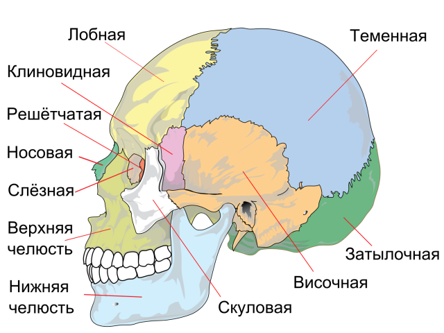
Anatomy - the structure and functions of the human skull: a scheme with a description
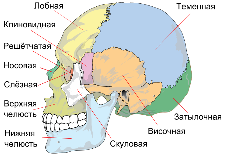
The human skull consists of a bone frame. He, in turn, includes 23 bones. They are needed to protect the brain from damage. Total 7 unpaired, and 8 paired bones. Here is a detailed structure of the skull - a scheme with a description, view of the side:
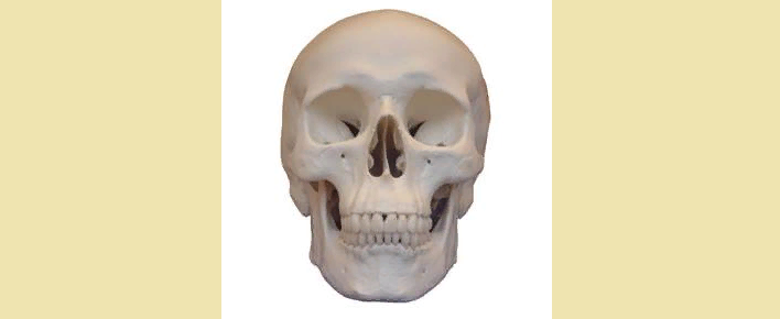
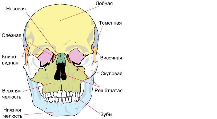
Here is a description scheme - front view:
The human skull is an integral part of the musculoskeletal system. It has two main sections (facial, brain). Each department has a strong effect on the human body as a whole.
The functions of a person’s skull are important for the life of the whole organism. It is necessary to implement several tasks at once:
- Serves as a strong frame for the human brain, nasal sinuses and eye sockets.
- Promotes the combination of facial muscles, chewing muscles.
- Actively participates in speech.
- Helps chew food, i.e. It is necessary for normal digestion of a person.
Below you will find a detailed description of the name of the bones and joints of the skull. Read further.
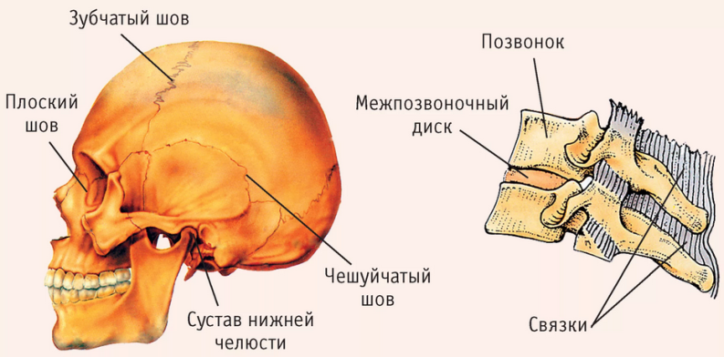
The seams of the skull of man
The seams of the skull are at the base of the skull and are continuous bone connections. The seams in an adult and baby differ. In a mature person, these are fibrous compounds, in a baby - inter -cell membranes.
There are still not permanent or temporary seams. They are formed due to fusion or not fraining several ossification places. For example, two parts of the frontal bone did not grow together. In such a situation, the sagittal seam originates from Glabella, maybe even a little higher.
If there is an inter -mobile or cutting bone, then the seam will be cutter. When the parietal bone doubled - an intermediary seam.
In most cases, together with human growing up - cartilage seams are delayed in full or partially. Then the connective tissue plate will be replaced with bone tissue. Cartilage compounds are formed from fibrous cartilage and are located at the base of the skull. Over time, the cartilage is replaced by bone. Therefore, synostosis is formed on the wedge-and-white synchondrosis by 25 years.
Differences in the skull of a newborn and an adult, female from male
Differences in the skull of a newborn and an adult, female from male
There are great differences between the structure of the head of the newly born child and an adult, and quite significant:
- The newborn has a more developed brain zone, and the front is much less developed. The front department of the skull begins to actively form with 13 years of age.
- Newborn children have fontanels on the head. Fines are the remains of a membranous skull and they are located at the site of the joint of the seams among themselves. Therefore, in this part, the pulsation of the artery of the brain is so clearly visible.
- The arch of the skull, the newly born baby, is well developed, in contrast to the base. There are no seams between the cranial bones of the newborn, so the bones are more plastic than that of an adult.
- The eye hole in newborns is much larger than that of an adult.
- The kid was only born, so the jaw is poorly developed. Based on this, we can conclude that the facial zone is less high.
- There are no protrusions, tubercles on the child’s face, i.e. Poles of muscle connection are poorly visible.
The nasal cavity is very small, poorly developed. Because of this, children have breathing problems. - The auditory holes are very large - this is one of the reasons why young children get otitis media.
The difference in the structure of the skull in a man and a woman, first of all, is in size. As a rule, a man has much more. Here are the differences between the female skull from the male:
- The male skull box is more angular than female.
- The places of attachment of vertebrae, chewing muscles are strongly expressed on it.
- The woman has a more even, smooth skull.
- In the stronger sex, the superfluous arcs and the nose of the nose are well developed, and in women, parietal tubercles are better developed.
- The lower part of the face (lower jaw) is much more developed in men.
It is uneven, rough, clearly visible places of fastening of the porch muscles.
What parts is the brain skull divided into?

The human skull is divided into two main parts - facial and brain.
Bones of the brain consist of:
- Occipital
- Wedge -shaped
- Lattice
- Frontal
- Temporal
The bones of the front part:
Unpaired:
- Soshnik
- Lower jaw
- Hand -language bone
Paired:
- Upper jaw
- The lower nasal sink
- The palatine
- Sculpture
- The nasal
- Tearful
The cranial bones are connected to each other and are in a stationary state. An exception is the lower jaw. There are joints and a sub -language bone in it, which is also constantly moving.
The front and lateral norms of the skull of a person
The facial norm consists of the frontal department, eye sockets, pear -shaped opening, upper jaw, teeth and chin protrusion. This projection helps to study the front of the human skull in great detail.
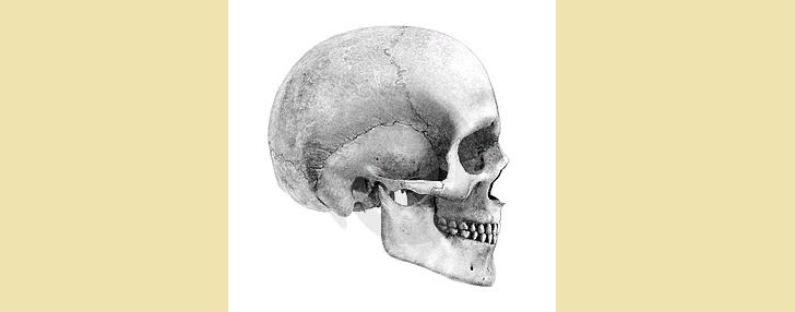
The second norm is lateral. She has another name - lateral. The most in detail considers the ratio of the departments of the skull - brain, facial, vault and base, their interaction. If you study a more detailed model, then a skeleton of the head, a temporal pit, a hearing hole and a shameless process are distinguished in it.
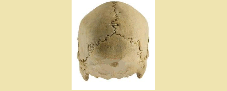
The occipital rate of the skull of a person
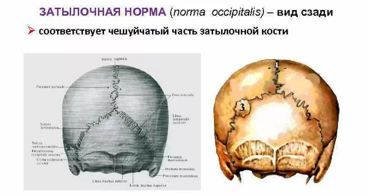
The occipital rate is represented by the back of the base of the human skull. In this norm, there is a lambdrod and torn-and-consuming seam. Also, such a norm is studied by a special process and occipital elevation.
The front skull
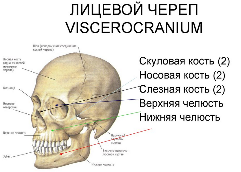
The front skull includes 9 bones: 6 - paired and 3 - no. The lower jaw and the zygomatic bone determine the shape of a person's face.
Cheekbone:
- It is quite asymmetric, irregular in shape.
- It includes wide plates and a little spongy.
- Refers to the side department.
Nebena bone:
- It is steam room and is located next to the upper jaw and a winged process.
- The palate helps to form the walls of the oral cavity.
- It consists of 2 plates (horizontal and vertical).
Labbly bone:
- This is the smallest bone in the facial department.
- She is almost transparent, she has four sides.
- The lacrimal bone looks like a small plate.
- It is located in the eye socket between the frontal process and the eye plate.
Nasal bone:
- This is a long four -sided plate.
- It is she who forms the back of the nose.
- The outer surface is flat, and the entire inner surface is covered with furrows.
- The lower nasal sink is quite thin, slightly curved. The edges of the bone are elongated.
Soshnik:
- This is a sufficient thin bone, it also has four sides.
- It forms the lower back of the nasal septum.
- Outwardly, the bone looks like a pair of plates that are connected below.
Hand -language bone:
- This is not one of the bones of the skull, but is studied in a group with them, because it is close to them very much in development.
- Located in the cervical region, a strongly curved plate.
- The outer side is uneven, the muscles that are located in the neck are attached in it.
- The inside, on the contrary, is very smooth.
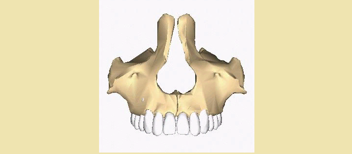
Upper jaw:
- The largest part of the face.
- It is located in the middle, so it adjoins all the other.
- It immediately forms the eye, oral and frontal parts.
- Although the jaw is large, but very light, because inside it there are sections filled with air.
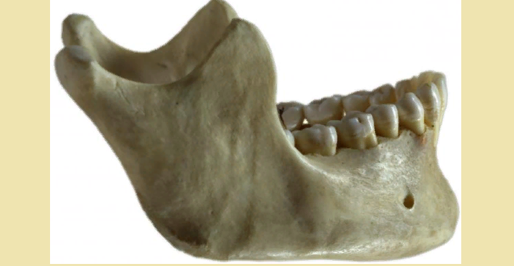
Lower jaw:
- Very strong bone of the facial department.
- It is attached to the temporal part and forms an arc.
This is the only joint that can move.
Brain skull
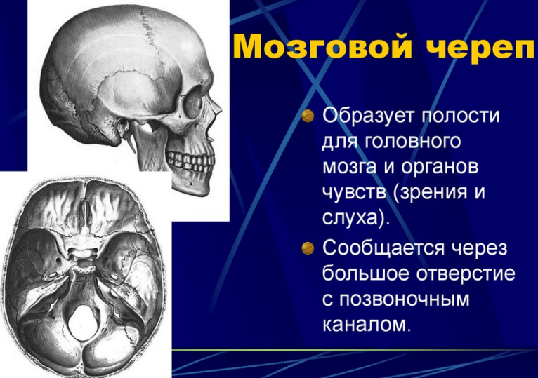
The brain department is the main part of the structure of the human skull. Its structure looks like:
Occipital bone:
- It consists of two lateral parts, scales and the main part.
- The main part is connected to the wedge -shaped bone, and they form a ramp.
- On the scales, its outer part there is an occipital tubercle.
Sphenoid bone:
- It includes 6 processes.
- Boles form branches - these are large wings, small wings and porcaric processes.
- On the outer surface of the body there is a pituitary gland.
Ethmoid bone:
- It is formed from a horizontal plate and perpendicular, a pair of orbital plates, a pair of labyrinths.
- Labyrinths are small cells that are separated from each other with plates.
Frontal part:
- It includes scales, a couple of eye departments and nasal sinuses.
- The eye departments slowly flow into the supra -ironic edges.
- The air sinus is divided into two parts with a partition.
- On the scales of the frontal bone there are frontal bugrs and super -term arcs.
Temporal bone:
- It contains a hearing aid, a sleepy artery.
- There is a auditory pass on the outer part, in the front there is a special auditory pit.
- The zygomatic process begins from the scales and goes to the process of the lower jaw, forming the zygomatic arc.
Brain skullit has a larger size than the front skeleton. In addition, it carries out one of the most important functions - protecting the brain from external damage. Although the bones in it are much smaller than in another department, they are more durable and thick.
Skull from the inside: Scheme
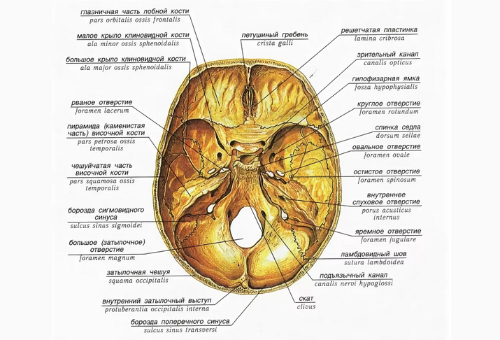
The inner surface of the skull is curved, quite tuberous and uneven. It completely repeats the outline of the base of the brain. It contains a large amount of veins, various arteries. The skull inside consists of three holes:
- Front. Forms the eye zone. It is extremely uneven - there are elevations and hollows, many fibers pass through the front.
- Average. It is located deeper than the first. It consists of two parts: the main and two side.
- The back. It is even deeper. It is located in the occipital part, in the middle there is a small hole.
The internal structure of the skull is described in detail above the diagram.
Racial features of the structure of the human skull
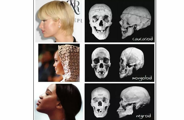
The skulls of people can have differences and racial features that every race has. For example:
- Europeans have a very expressive nose. It has a high nose bridge, narrow with a pronounced protrusion. There are features in the bite and planting of teeth. In Europeans, the teeth are located vertically, the fangs are deeply planted. The cheekbones of this race are generally invisible.
- Representatives of the Mongoloid race, on the contrary, have pronounced cheekbones. The shape of the face is flat, elongated. The ledge of the nose, unlike Europeans, is very small.
- The Australo-Neogroids have crooked teeth, a face with a straight high forehead.
Features of the structure of a particular race - do not give anyone the advantages in the struggle for survival, do not affect the viability of the population. Although German scientists in the 40s refuted this theory. They divided the population into two races: the highest and lower - depending on the structure of the skull.
It was believed that people of the northern race are more viable, healthy and smart, therefore they should lead the rest - less successful races. Later, scientists proved that all races can have an elongated face and overhanging eyebrows (fascist Germany believed that these are signs of only the northern race). The theory of the superiority of any races and nations over others turned out to be non-viable.
Sexual differences in the structure of the skull
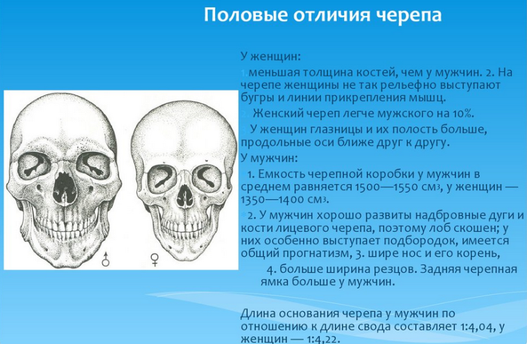
Skulls may differ among themselves not only because of the race to which a person belongs. The difference in the structure of the skull will be noticeable between people of different sexes. Here are the gender differences in the structure of the skull:
- The difference in the structure of the skull in a man and a woman, first of all, is in size. As a rule, in a man it is much larger and harder.
- The male skull box is more angular than female. The places of attachment of vertebrae, chewing muscles are strongly expressed on it. The woman has a more even, smooth skull.
- In the stronger sex, the superfluous arcs and the nose of the nose are well developed, and in women, parietal tubercles are better developed.
- The lower part of the face is much more developed in men. It is uneven, rough, clearly visible places of fastening of the porch muscles.
- At the stronger sex, the forehead is a little as if mowed, and the number of a more rounded shape. Often in this zone there are small cones, hills. In women, the theme, on the contrary, is flat and the forehead is vertical.
- In men, the front part is much better developed. It is more powerful and more than female.
- The transition between the nose and forehead of the stronger sex is clearly visible, and in women it is much less noticeable.
- The eyes of men are planted below, have the shape of a rectangle, the upper part is wider. In girls, the eyes are located above, a softer round shape, there is no thickening in the upper part.
These differences between the structure of the skull of a man and a woman do not affect, do not affect the ability of survival, or on the mental abilities of people. This is only a reflection of the process of evolution and biological features of the structure of the skeleton and the functioning of the body.
Video: 3D anatomy of the skull. Types of compounds of the bones of the skull
Read on the topic: