This article describes the types of human joints, as well as types of development and symptoms of the most common disease.
Content
- What are the types of human joints, bone joints: joint structure, main elements, functions, description scheme, table
- Types of development of hip joints by column, by ultrasound: Description
- Types of development of hip joints in newborns, infants: Features
- Types of physiological immaturity of joints 1A, 1B, 2, dysplasia: what does this mean?
- Type - knee joint: Anatomy
- Type - shoulder joint: structure
- Type - ankle joint: Anatomy
- Type - elbow joint: structure
- What types of joints are joints?
- Types of joint movement
- Types of arthrosis of the joints: symptoms of the disease
- What type of collagen is needed for joints with arthrosis?
- Joints in type 2 diabetes: what's the danger?
- MRI of the knee joint of the open type: how it goes, video
- Video: MRI of the knee joint
- Video: General arthrology. Connections of the bones of the skeleton of the body
The joints play an important role in the body. A simple person knows a little about these types of bone connection. If you are studying at a medical institute or college, then you need to know more. In this article we will consider what types of joints exist in an adult, in a newborn. You will also learn about the joints in diabetes, which you need to beware and so on. Read below.
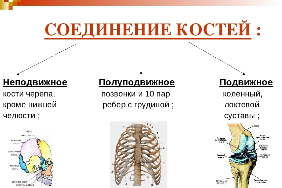
What are the types of human joints, bone joints: joint structure, main elements, functions, description scheme, table
Below in the picture you will see what types of human joints, bone joints, as well as their descriptions.

The structure of the joint is simple and it is easy to remember. Here are the main elements and a description scheme:

Here are the tables of the structure, the names of the joints with their functions:


Types of development of hip joints by column, by ultrasound: Description
In 1980, one famous scientist R. Count developed a unique screening methodology, with which you can determine the type of joint development by ultrasound. To confirm the diagnosis, functional tests are performed along with ultrasound. Below you will see a photo of an ultrasound of the hip joint. It marks the angles (A and B), with which the type of development of the Tasobedr is performed. joint according to the graph methodology.

With help angle a You can evaluate the development of the bones of the acetabulum. By corner b It can be determined how the development of the cartilage of the swivel zone occurs. The smaller will be angle a And more angle b, the greater the degree of underdevelopment of the joint. The description of the types of development is lower. Read further.
Types of development of hip joints in newborns, infants: Features
In newborns, the structure of the hip joints differs from the structure of these parts of the body in adults. The fact is that their significant departments consist of cartilage fabric. The ossification of the femur begins to occur approximately in 8 weeks embryogenesis. Ossamation is the development of bones, their formation, ossification. And embryogenesis is the process of developing the embryo.
- In the baby, the ossification nucleus in the body develop in four, five and six months in the intrauterine state.
- When the child is born, many departments of the pelvic bones still retain their cartilaginous structure.
- In their place, a layer of cartilage, which is called in the medical language, continues to remain in their place Y-shaped cartilage.
Unfortunately, bones and cartilage are not always in the right position.
- Normally in 3-4 months The child, the location of these joints should be about 25-30 degrees.
- AT 5 months-2 years – 20 - 25 degrees.
- AT 2-3 years - From 18 to 23 degrees.
- If the angle of "inclination" is increased, we can talk about subluxation, dislocation, high dislocation and dysplasia.
What does it mean:
- With subluxation in infants, the angle can be up to 35 degrees.
- With the dislocation, the numbers are higher - up to 40 degrees.
- With high dislocation and dysplasia, the indicators will be greater 40 degrees.
Deviations in the development of the motor apparatus in infants in the early stages are determined using ultrasound. Usually the result is unmistakable and ultrasound helps to determine existing problems. In addition, this type of study is harmless to the life and health of a small child.
It is worth knowing: If the baby has dysplasia in which the joints develop incorrectly, then the orthopedic doctor can determine this without an ultra-sound study, but ultrasound will only confirm such a diagnosis.
Types of physiological immaturity of joints 1A, 1B, 2, dysplasia: what does this mean?

There are several types of types of hip joints in newborn, babies. Here are the types of physiological immaturity of the joints and what it means:
- Type IA: This is a normal mature joint, observation is not required in the future.
- Type 1b: Close to normal, repeated ultrasound in this case is prescribed after 3 months.
- The second type is II. It has subgroups A, b, C and D. If the doctor observes this type of joint development of the baby, then you need to do a second ultrasound every 1-3 months.
Read more:
In the first group (1a, 1b) - You need to do only ultrasound.
- The doctor will observe development and if everything is fine, after a year, the ultrasound is performed only 1 time in year.
- If the disease is classified as subgroup b, then we can safely talk about joint dysplasia. Need Pavlik’s stirrup and control ultrasound 1 time per month.
Development of the joint of the type II with, then it is already about severe joint dysplasia, the bondage, treatment is necessary.
- May need a gypsum bandage for 3 weeks, and then stirrup. Ultrasound control is made 1 time per month.
If the child type II D, Doctors will already talk about severe dysplasia, a bondage close to the decentration of the head.
- A gypsum bandage is needed for 3 weeks. After that, the child will wear Pavlik’s stirrups and it will be necessary to do an ultrasound 1 time per month.
The third type – III. There are two subspecies.
- In the first group - severe dysplasia and subluxation. We need a closed reposition and a gypsum bandage, the stirrups of Pavlik, ultrasound control every month.
- In the second group - cases of serious damage, chronic thigh dislocation is possible.
Fourth type - IV.
- With him, the alpha-corner is less 43 degrees.
- The bone part is almost flat, the joint lip is clamped.
- Such children urgently require treatment. Ultrasound control 1 time per month.
If a problem is detected, it needs to be treated. Modern methods allow you to achieve good results.
Type - knee joint: Anatomy
The knee joint refers to the movable type of joints, the category is block. It has one axis of movement, which passes along the entire length of the joint. Here is the anatomy of the knee joint:

The ligament helps stabilization of the joint and prevents the shift of the lower leg. Therefore, it is considered one of the most important elements of the knee joint. This joint has a complex structure (this confirms the picture above). He helps a person walk, perform physical activity, but this is the place that is most often damaged.
Type - shoulder joint: structure
A special anatomy of the shoulder joint helps the hands be mobile. It consists of cartilage, ligaments and muscles. Here is a structure of this type of human joint as the shoulder joint:

The shoulder blade when the hand moves almost always remains motionless. The diameter of the head of the shoulder is three times larger than its "bed". These different sizes help make the amplitude of the movements of large and wide. So that the head does not go beyond the hollow, the joints surround the ligaments and muscles.


Type - ankle joint: Anatomy
The ankle joint is one of the most important joints. It consists of muscles, ligaments, cartilage and bones. With the coordinated work of all these elements, the foot of the leg can move in different directions, and a person can walk. Here is an anatomy of the ankle type of joint:
Bones.

Muscles.

Blues.

Type - elbow joint: structure
The elbow joint consists of three bones: shoulder, elbow, radiation. Between themselves, they are connected by articular cartilage. Around the joint, ligaments and muscles pass, which warn this element from fractures and displacements. Here is the structure of the elbow type of joint:

Here are more the anatomy of the bones and ligaments of the elbow joint:

The muscles of the elbow joint are a complex structure. Here is the structure of the muscle frame in this area:

What types of joints are joints?
There are different types of bones of human bones. They help bone perform various functions and provide mobility. There is no single classification of types of compounds. They are divided into mobile and motionless. There is also a third type of compound - these are semi -navigable joints.
Joints are mobile types of joints. Although there are also cartilage and other connective tissue in the semi -adoles of bone joints. Therefore, this species can also be called joints.
Types of joint movement
Bone compounds in the body are characterized by various values, mechanisms and configuration. In addition to them, the joint movement is regulated by its axes:
- Frontal and sagittal axis They are part of the planes dividing the body into four parts. The front plane on the front and rear, and the sagittal - on the left and right parts.
- Vertical axis - This is a personal axis of the body.
In total, several ways to change the position of the joint relative to the axis are distinguished:
- Adduction (approximation to the center) and abduction (removal from the center) - characterized by a movement relative to the sagittal line.
- Flexion and extension - characteristic of the movement along the front axis.
- Rotation It has three directions: rotation inward (pronation), rotation from the inside (supination) and conical rotation. This is a method of joint movement of the vertical axis.
The joint is based on a geometric figure, so the joints acquire the possibility of moving along the axes, from the only one to all possible, thanks to the properties of these figures.
Types of arthrosis of the joints: symptoms of the disease

Joints in type 2 diabetes: what's the danger?

Changes in the joints in patients with type 2 diabetes, occur in a large number of cases. Especially in severe diabetes, inflammatory processes occur, which lead to degenerative disorders in the joints. What is the danger of joint damage in type 2 diabetes? Here are the answers:
- Arthritis It can occur in the knee and hip joints, and significantly violate their functions.
- In severe forms of arthritis and arthrosis, may need surgery to replace the affected joint.
- With osteochondrosis Degenerative changes appear in different parts of the spine. Their trophy is violated, and the destruction of the vertebrae begins. Because of which there are painful sensations, arrhythmia, headaches, dysfunction of the gastric and intestinal tract. The functionality of patients is limited.
- Diabetic hyropathy can develop (defeat of the hands). This leads to the difficulty of extension and bending of the joints of the hands. She can negatively affect the quality of life of the patient.
In addition, there is a tendency to develop osteoarthritis and osteoporosis. The bones become more fragile, which often leads to fractures. The recovery period is delayed due to the increased blood sugar.
MRI of the knee joint of the open type: how it goes, video
Magnetic resonance imaging (MRI) of the knee of the open type is a common study, the purpose of which is to identify changes in the condition of tissues and organs of the patient. MRI is the most modern type of study, in connection with which it has fewer contraindications and allows you to examine an organ that is exciting patient in more detail.
Preparation for MRI:
- Magnetic waves in the MRI implies that the patient should not have metal objects during the study (jewelry, prostheses, etc.).
- The presence of non -removable metal prostheses or pins at the site of the examination is a contraindication.
How MRI goes:
- The patient is placed in an open -type tomograph, after which a strong magnetic field is created in it.
- Then the fluctuations in the studied area are transmitted to the computer.
- The procedure itself occupies 15-20 minutes And it is very important that the patient retains immobility during the entire procedure, so that the final result is more accurate.
- In some cases, during the examination, a contrast medium is introduced into the knee joint.
Here is a video that shows how an MRI of the knee joint goes:
Video: MRI of the knee joint
Human joints are complex elements that help us move. From this article you learned about the structure of these elements, the symptoms of the most common disease and diagnostic methods.