All about planned and additional ultrasound during pregnancy. What parameters are evaluated and how the fetus should develop in weeks.
Content
- First ultrasound during pregnancy: for what time?
- Is there a dangerous ultrasound in the early stages of pregnancy?
- Will an early pregnancy be determined for ultrasound?
- Fetal ultrasound standards: decryption table
- At what period does the ultrasound determine the gender of the child?
- Planned and additional ultrasound: Indications. When is the ultrasound during pregnancy in trimester?
- How long is the first ultrasound?
- At what period of pregnancy is the second ultrasound do?
- Dimensions of the fetus for weeks by ultrasound: Table
- At what period of pregnancy do the third ultrasound do?
- How much ultrasound do you need to do during pregnancy?
- Video: All about ultrasound during pregnancy
- Video: planned ultrasound
For future parents, an ultrasound is always both a joyful and anxious event. Indeed, on the one hand, the examination makes it possible to get acquainted with the baby, on the other, this is still a medical examination to identify possible pathologies.
First ultrasound during pregnancy: for what time?
In accordance with medical standards approved by WHO, the first ultrasound must be done at 11-14 weeks. As a rule, doctors prescribe it at 12 weeks. There are several reasons at once, why ultrasound must be carried out precisely during this period:
- Only in this time period can the presence of Down syndrome and some other severe pathologies be excluded, measuring the thickness of the collar space (a tubercle in the crown and neck, which the fetus has in this period)
- Only up to 15 weeks can you set the gestational age with high accuracy. After 15 weeks, genetic factors begin to influence the size of the fetus, but until that time they all develop almost the same
On the first ultrasound, as a rule, it is still impossible to determine who the boy or girl is born. But it will be possible to compare the norms of ultrasound of the fetus recorded in the literature with the image on the monitor and hear the heartbeat of the child.

Is there a dangerous ultrasound in the early stages of pregnancy?
It can be completely confidently argued that there was not a single study that would confirm that an ultrasound is harmful. In the world, even one fact is not recorded that would connect ultrasound with developmental abnormalities.
But the exact and justified from a scientific point of view, the answer to this question does not exist. Large -scale studies on this subject simply were not carried out, probably for the same reasons why the effect of drugs on pregnancy was not checked. No one will give permission to conduct such experiments.
Nevertheless, there are data confirming that high doses of ultrasonic irradiation slowed down the course of pregnancy in animals. In addition, it is known that the vital indicators and norms of ultrasound of the fetus when they make ultrasound change, the heartbeat quickens, the child becomes more mobile, which means that children feel the effect of ultrasound.

It is believed that it is not necessary to carry out ultrasound diagnostics too often in the first trimester, and there is a logical justification for this. Ultrasound is waves that cause cell fluctuations and their heating.
The smaller the size of the fetus, the larger the effect on it and vice versa, the larger the gestational age, the less ultrasound can affect the child. If you add up all opinions, it turns out that it is better to minimize the amount of ultrasound, but if there are medical indications for additional examinations, then they must definitely be done.

Will an early pregnancy be determined for ultrasound?
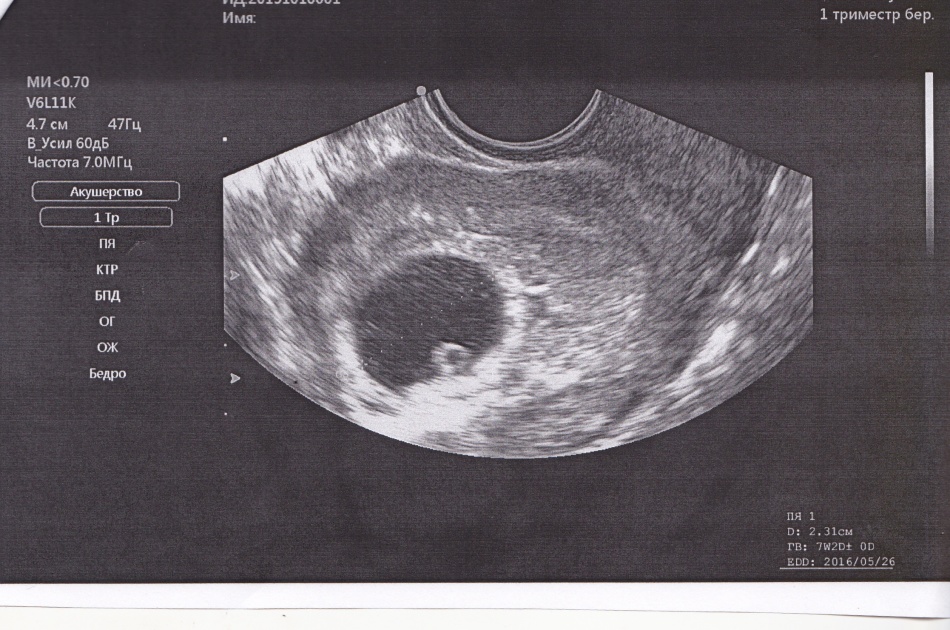
It is possible to detect a fetal egg, starting from a period of 5 obstetric weeks, after 7 days of delay in menstruation. Be prepared for the fact that a vaginal sensor will be used for the examination. This type of ultrasound is shown up to 11 weeks, since an ultrasound of an ordinary sensor through the abdominal wall will be uninformative.

Fetal ultrasound standards: decryption table
Doctors of the diagnosis ultrasound dedicated the standard size of the fetus by ultrasound weeks, and the size of the embryo is extremely accurately determined by the gestational age and the date of conception. Until 14 weeks, the size is characterized by such a parameter as KTR (coccyge-dark size), that is, the length from the coccyx to the crown. Using the fetal ultrasound table, you can compare the size of the fetus and the number of obstetric weeks.
| Weeks and days | KTR (mm) | Weeks and days | KTR (mm) |
| 6+3 | 7 | 10+3 | 36 |
| 6+4 | 8 | 10+4 | 37 |
| 6+6 | 9 | 10+5 | 38 |
| 7 | 10 | 10+6 | 39 |
| 7+2 | 11 | 11 | 40-41 |
| 7+3 | 12 | 11+1 | 42 |
| 7+4 | 13 | 11+2 | 43-44 |
| 7+5 | 14 | 11+3 | 45-46 |
| 7+6 | 15 | 11+4 | 47 |
| 8 | 16 | 11+5 | 48-49 |
| 8+1 | 17 | 11+6 | 50-51 |
| 8+2 | 18 | 12 | 52 |
| 8+3 | 19 | 12+1 | 53 |
| 8+4 | 20 | 12+2 | 54-57 |
| 8+5 | 21 | 12+3 | 58 |
| 8+6 | 22 | 12+4 | 60-61 |
| 9 | 23 | 12+5 | 62-63 |
| 9+1 | 24 | 12+6 | 64-65 |
| 9+2 | 25 | 13 | 66 |
| 9+3 | 26-27 | 13+1 | 68-69 |
| 9+4 | 28 | 13+2 | 70-71 |
| 9+5 | 29 | 13+3 | 72-73 |
| 9+6 | 30 | 13+4 | 75 |
| 10 | 31-32 | 13+5 | 76-77 |
| 10+1 | 33 | 13+6 | 79-80 |
| 10+2 | 34-35 | — | — |
If the deadlines and size do not match, do not be upset, the discrepancy up to 3 days is considered permissible. In addition, for the calculation, the standard period of ovulation is taken, and in practice it could occur earlier or later, errors during the study itself are possible.

At what period does the ultrasound determine the gender of the child?
As a rule, the gender of the child is determined between 20 and 24 weeks, on the second planned ultrasound. Sometimes the floor is able to determine at 13 weeks, but this requires a number of conditions:
- the presence of an experienced specialist
- high-quality ultrasound diagnosis
- the right position of the fetus.
Often, when determining the floor, errors occur: the edema of the labia that happens in girls can be mistaken for the penis, and the boy who tightly squeezes the legs can be erroneously classified as a girl. Therefore, the gender of the baby still remains a mystery, until the visit to the hospital and his birth.

Planned and additional ultrasound: Indications. When is the ultrasound during pregnancy in trimester?
Many expectant mothers are interested in the question: how much ultrasound should be done during pregnancy? The answer is simple: as much as necessary, but there are at least three mandatory ultrasound, one for each trimester.
Additional studies are prescribed for any period if there are anxiety symptoms or if, upon a planned examination, suspicions of pathology have occurred.
Often unscheduled examinations are prescribed in the late stages to make sure that a woman will not have problems during natural birth.

How long is the first ultrasound?
You need to go to your first ultrasound no later than 11-14 weeks. Sometimes the first ultrasound is shown much earlier, it is done to establish the very fact of the presence of a uterine developing pregnancy. However, it is not worth passing it without the direction of the doctor, there is a clear list of indications to send the patient for ultrasound up to 11 weeks, this is: this:
- Bloody discharge, which indicates the threat of termination of pregnancy
- Non -compliance with the size of the uterus of pregnancy
- Conducting artificial fertilization (IVF) or the use of other techniques stimulating conception
- Problems with bearing in the past
- Pain in the lower abdomen
Please note that pain in the lower abdomen is a fairly ambiguous symptom. Sometimes they signal such a dangerous pathology as an ectopic pregnancy. But more often the reason is more banal: pregnant women have a tendency to constipation and bloating and, it is possible to get rid of pain, it is enough to reconsider your nutrition. In summer, for the prevention of constipation, add plums or other fruits with a dense peel to your diet, in winter - kiwi are perfect.
In addition, minor painful sensations are natural, the body is prepared for pregnancy and the ligaments are stretched. But these pains should be short -lived, barely tangible and not to have a clear localization. With an ectopic pregnancy, the pain is localized in one place, has a pulling character and increases over time.

At what period of pregnancy is the second ultrasound do?
The second ultrasound study is shown from 20 to 24 obstetric weeks. In practice, it is usually prescribed at 21 weeks. At this time, there is already the opportunity to determine the sexual belonging of the unborn child, the main internal organs are formed and clearly visible at this time, so possible pathologies are visible.
Dimensions of the fetus for weeks by ultrasound: Table
In addition to internal organs, limbs are studied, and their length is measured. Attention is also paid to the number of amniotic fluid, placenta and umbilical circulation. The standard weight and other norms of the fetal ultrasound during pregnancy in trimester are given in the picture in the table below.
| A week | 11 | 12 | 13 | 14 | 15 | 16 | 17 | 18 | 19 | 20 |
| Growth | 6,8 | 8,2 | 10 | 12,3 | 14,2 | 16,4 | 18 | 20,3 | 22,1 | 24,1 |
| The weight | 11 | 19 | 31 | 52 | 77 | 118 | 160 | 217 | 270 | 345 |
| BRG | 18 | 21 | 24 | 28 | 32 | 35 | 39 | 42 | 44 | 47 |
| DB | 7 | 9 | 12 | 16 | 19 | 22 | 24 | 28 | 31 | 34 |
| DGK | 20 | 24 | 24 | 26 | 28 | 34 | 38 | 41 | 44 | 48 |
| A week | 21 | 22 | 23 | 24 | 25 | 26 | 27 | 28 | 29 | 30 |
| Growth | 25,9 | 27,8 | 29,7 | 31,2 | 32,4 | 33,9 | 35,5 | 37,2 | 38,6 | 39,9 |
| The weight | 416 | 506 | 607 | 733 | 844 | 969 | 1135 | 1319 | 1482 | 1636 |
| BRG | 50 | 53 | 56 | 60 | 63 | 66 | 69 | 73 | 76 | 78 |
| DB | 37 | 40 | 43 | 46 | 48 | 51 | 53 | 55 | 57 | 59 |
| DGK | 50 | 53 | 56 | 59 | 62 | 64 | 69 | 73 | 76 | 79 |
| A week | 31 | 32 | 33 | 34 | 35 | 36 | 37 | 38 | 39 | 40 |
| Growth | 41,1 | 42,3 | 43,6 | 44,5 | 45,4 | 46,6 | 47,9 | 49,0 | 50,2 | 51,3 |
| The weight | 1779 | 1930 | 2088 | 2248 | 2414 | 2612 | 2820 | 2992 | 3170 | 3373 |
| BRG | 80 | 82 | 84 | 86 | 88 | 89,5 | 91 | 92 | 93 | 94,5 |
| DB | 61 | 63 | 65 | 66 | 67 | 69 | 71 | 73 | 75 | 77 |
| DGK | 81 | 83 | 85 | 88 | 91 | 94 | 97 | 99 | 101 | 103 |
BRG is a biparietal head size. DB - hip length. DGK - chest diameter
At what period of pregnancy do the third ultrasound do?
The third ultrasound must be passed 32-34 weeks or earlier if there is good reason to believe that there will be premature birth. Its main task is to determine whether natural birth is possible.
The arrangement of the fetus and the location of the placenta are considered, the umbilical cord is excluded and the size of the child’s head is measured.

How much ultrasound do you need to do during pregnancy?
The first apparatus for ultrasound appeared more than 50 years ago. Now this research method is considered the safest for pregnant women and is widely used all over the world. Ultrasound not only makes it possible to get objective information, but also helps to dispel the anxieties of future parents. Therefore, the number of procedures will depend on the development of the fetus and health of the mother and is provided individually for each pregnant woman.
Video: All about ultrasound during pregnancy
Acusher-gynecologist and an ultrasonic diagnostic specialist, on the appropriateness of ultrasound in certain cases