

In this article, an anatomical skeleton of the legs, feet, arms, brushes, pelvis, chest, neck, skull, shoulder and forearm of a person will be considered: scheme, structure, description.
Content
- Human skeleton - bones, their structure and names: scheme, photo, from the side, from the back, description
- The main parts of the human skeleton, quantity, bone weight
- Anatomical skeleton of the legs, foot of a person: scheme, description
- Anatomical skeleton of the arm, human hand: scheme, description
- Anatomical skeleton of the shoulder and forearm of a person: scheme, description
- Anatomical skeleton of the chest, human pelvis: scheme, description
- Anatomical skeleton of the neck, human skull: scheme, description
- What is the role of a person’s skeleton, which ensures mobility, which belongs to the mechanical function of the bones of the skeleton?
- Human skeleton bones: What are interconnected?
- What are the features of the structure of the human skeleton associated with directness?
- What is the longest, massive, strong and small bone in a human skeleton?
- What bones are tubular in a human skeleton?
- What bones in a person’s skeleton are connected mobile with the help of a joint and motionless?
- What fabric is the basis of the bones of the skeleton, what substance gives a person’s skeleton, what is the composition of the bones?
- How old is the skeleton of a person?
- Video: Human skeleton, its structure and meaning
The skeleton is the supporting support of organs and muscles that provide our life, and makes it possible to move. Each part of it consists of several departments, and they, in turn, are of bones that are able to change with time and subsequently injuries.
Sometimes there are anomalies from the side of bone growth, but with proper and timely correction, they can be put in an anatomical form. In order to identify development pathologies in time and provide first aid, you need to know the structure of the body. Today we will talk about the structure of the human skeleton, in order to once and for all to understand the whole variety of bones and their functions.
Human skeleton - bones, their structure and names: scheme, photo, from the side, from the back, description
The skeleton is the totality of all bones. Each of them also has a name. They differ in structure, density, shape and different purpose.
Having been born, the newborn has 270 bones, but under the influence of time they begin to develop, united among themselves. Therefore, there are only 200 bones in the body. The skeleton has 2 main groups:
- Ossial
- Additional
In the first category, the following groups of bones are located:
- Skull (facial, brain part)
- The chest (includes 12 vertebrae of the thoracic region, 12 pairs of ribs, sternum and its handle)
- Spine (cervical and lumbar)
The additional part includes:
- The belt of the upper extremities (including collarbone and shoulder blades)
- Upper limbs (shoulders, forearms, brushes, phalanxes)
- Belt of the lower extremities (sacrum, tailbone, pelvis, radiation bone)
- The lower limbs (patella, femoral, tibia and fibulas, phalanxes, foresaw and plus)
Also, each of the skeleton departments has its own nuances of the building. For example, the skull is divided into the following parts:
- Frontal
- Parietal
- The occipital
- Temporal
- Sculpture
- Lower jaw
- Upper jaw
- Lacrimal
- Nasal
- Reshet
- Cleem

The spine is a ridge that is formed thanks to the bones and cartilage built along the back. It serves as a kind of frame to which all other bones are attached. Unlike other departments and bones, the spine is characterized by a more complex placement and has several components of the vertebrae:
- Cervical department (7 vertebrae, C1 - C7);
- Thoracic region (12 vertebrae, Th1 - TH12);
- Lumbar region (5 vertebrae, L1 - L5);
- The sacral department (5 vertebrae, S1 - S5);
- The coccygeal department (3-5 vertebrae, CO1 - CO5).
All departments consist of several vertebrae that affect the internal organs, the possibility of the functioning of the limbs, neck and other parts of the body. Almost all bones in the body are interconnected, therefore, regular control and timely treatment are necessary for injuries in order to avoid complications in other parts of the body.

The main parts of the human skeleton, quantity, bone weight
The skeleton changes throughout a person’s life. This is due not only to natural growth, but also by aging, as well as some diseases.
- As mentioned earlier, at birth in a child there are 270 bones. But over time, many of them unite, forming a skeleton natural for adults. Therefore, fully formed people can have from 200 to 208 bones. 33 of them, as a rule, is not paired.
- The growth process can last up to 25 years, so the final structure of the body and bones can be seen in the X -ray picture upon reaching this age. That is why many people suffering from diseases of the spine and bones take drug treatment and various therapeutic methods only up to 25 years. After all, after stopping growth, the patient’s condition can be maintained, but it cannot be improved.
The weight of the skeleton is determined in the percentage of the total body weight:
- 14% in newborns and children
- 16% in women
- 18% in men
The average representative of the stronger sex has 14 kg of bones from the total weight. Women only 10 kg. But many of us are familiar with the phrase: "Wide bone." This means that their structure is slightly different, and the density is greater. In order to determine whether you are enough to use this type of people to use a centimeter, wrapping it around the wrist. If the volume reaches 19 cm or more, then your bones are really stronger and more.
Also affects the mass of the skeleton:
- Floor
- Age
- The weight
- Growth
- Nationality
Many representatives of different peoples of the world are significantly different from each other in growth and even physique. This is associated with evolutionary development, as well as a densely rooted genotype of the state.

In the main parts of the skeleton there are different amounts of bones, for example:
- 23 - in the cranial box
- 26 - in vertebrates
- 25 - in ribs and sternum
- 64 - in the upper limbs
- 62 - in the lower extremities
They are also able to change throughout a person’s life under the influence of the following factors:
- Childbirth
- Diseases of the musculoskeletal system, bones and joints
- Obesity
- Injuries
- Active sports and dancing
- Infutable nutrition
Anatomical skeleton of the legs, foot of a person: scheme, description
Legs belong to the lower extremities department. They have several departments and function thanks to mutual support.
The legs are attached to the belt of the lower extremities (pelvis), but not all of them are evenly placed. There are several that are located only from behind. If we consider the structure of the legs in front, then the presence of such bones can be noted:
- Femoral
- Flaspeches
- Boloversovs
- Nebertsovs
- Udlusny
- Plus
- Phalanxes
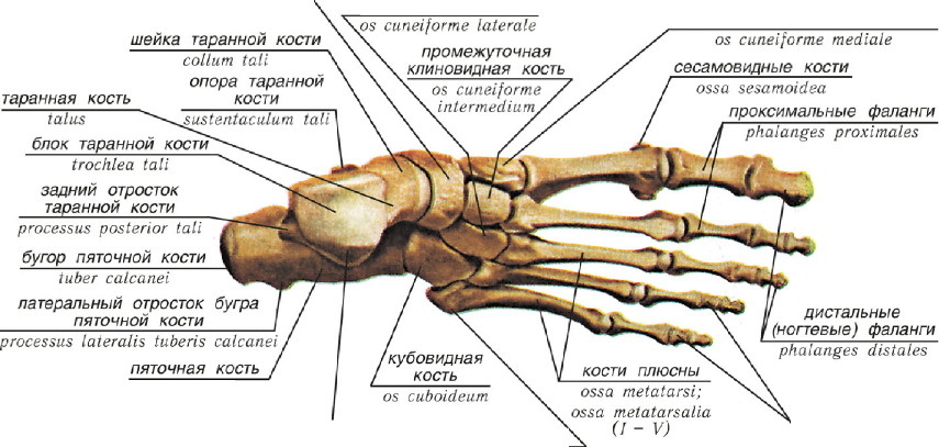
A heap bone is located behind. It connects the leg and foot. However, it is impossible to see her in the picture of an X -ray in front. In general, the foot differs in its structure and includes:
- Fifth bone
- Tarannoye
- Cube -shaped
- Scalp
- 3rd wedge-shaped
- 2nd wedge-shaped
- 1st wedge-shaped
- 1st plus
- 2nd plus
- 3rd metatarsal
- 4th plus
- 5th plus
- Basic phalanges
- End phalanx
All bones are interconnected, which allows the foot to function fully. In case of injury to one of the parts, the work of the entire department will be impaired, therefore, with various injuries, it is necessary to take a number of methods aimed at immobilizing the affected area and contact a traumatologist or surgeon.
Anatomical skeleton of the arm, human hand: scheme, description
Hands allow us to lead a full -fledged lifestyle. However, this is one of the most complex parts in the human body. After all, many bones complement each other's functions. Therefore, if one of them is damaged, we will not be able to return to the previous matters, without receiving medical care. The skeleton hands indicate:
- Collarbone
- Shoulder and blade joints
- Shoulder blade
- Shoulder bone
- The elbow joint
- Elbow bone
- Radiation bone
- Wrist
- Parlear bones
- The presence of proximal, intermediate and distal phalanxes

The joints connect the main bones among themselves, therefore they provide not only their movement, but also the work of the whole arm. Upon receipt of the injury of intermediate or distal phalanges, other sections of the skeleton will not suffer, since they are not connected to more important departments. But with problems with the collarbone, shoulder or elbow, a person will not be able to control and fully move his hand.
Therefore, if you have received any injury, you can not ignore the trip to the doctor, because in the case of fusion of tissues without proper help, this is fraught with complete immobility in the future.
Anatomical skeleton of the shoulder and forearm of a person: scheme, description
The shoulders not only connect the hands with the body, but also help the body to purchase the necessary proportionality from the point of view of aesthetics.
At the same time, it is one of the most vulnerable parts of the body. After all, the forearm and shoulders carry a huge load, both in everyday life and when playing sports with great weight. The structure of this part of the skeleton is as follows:
- Collarbone (has a connecting function of the shoulder blade and the main skeleton)
- Shoulder blade (unites the muscles of the back and arms)
- Cracious process (holds all the ligaments)
- Shoulder process (protects against damage)
- The articular hollow of the shoulder blade (also has a connecting function)
- The head of the humerus (forms the adjoining)
- Anatomical neck of the humerus (supports fibrous tissue of the joint bag)
- Shoulder bone (provides movement)

As you can see, all departments of the shoulder and forearms complement each other's functions, and are also placed in such a way as to protect the joints and thinner bones as much as possible. With their help, the hands move unhindered, starting from the phalanx of the fingers, and ending with collarbones.
Anatomical skeleton of the chest, human pelvis: scheme, description
The chest in the body protects the most important organs and spine from injuries, and also prevents their displacement and deformation. The pelvis plays the role of a frame, which preserves in a motionless state. It is also worth saying that our legs are attached to the pelvis.
The chest, or rather its frame consists of 4 parts:
- Two side sides
- Front
- Back
The frame of the human chest is represented by ribs, directly sternum, vertebrae and connecting their ligaments and joints.
The rear support is the spine, and the front of the chest consists of cartilage. In total, this part of the skeleton has 12 pairs of ribs (1 pair attached to the vertebra).
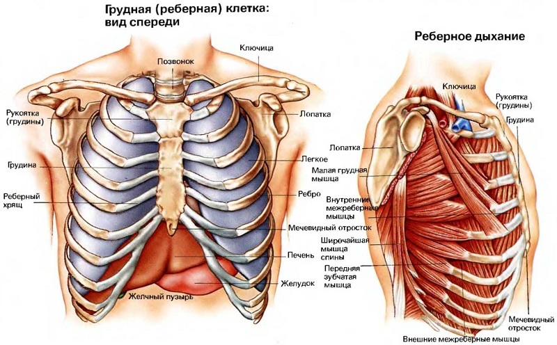
By the way, the chest wraves all vital organs:
- Heart
- Lungs
- Pancreas
- Part of the stomach
However, if diseases of the spine occur, as well as its deformation, ribs and parts of the cell can also change, creating excessive compression and pain.
The shape of the sternum may vary depending on genetics, type of breathing and general health. In infants, as a rule, the chest is protruding, but during the period of active growth it becomes not so visually expressed. It is also worth saying that in women it is more well developed and has advantages in width compared to the male.
The pelvis differs significantly depending on the human floor. Women are characterized by the following features:
- Large width
- Less length
- The shape of the cavity resembles a cylinder
- The entrance to the basin is rounded
- The sacrum is shortened and wide
- The wings of the iliac part are horizontally
- The angle of the pubic region reaches 90-100 degrees
The following characteristics are characteristic of men:
- The pelvis is narrower, but high
- The wings of the iliac part are located horizontally
- The sacrum is narrower and elongated
- Pubic angle of about 70-75 degrees
- Form of entrance "Card heart"
- Small pelvic cavity resembling a cone

The general structure includes:
- Large pelvis (fifth lumbar vertebra, the rear upper axis of the garter, sacral iliac art)
- Border line (sacrum, tailbone)
- Small pelvis (pubic symphysis, anterior upper part of the garter bone)
Anatomical skeleton of the neck, human skull: scheme, description
The neck and skull are complementary parts of the skeleton. After all, without each other they will not have mounts, which means they will not be able to function. The skull combines several parts. They are divided into subcategories:
- Frontal
- Parietal
- Occipital
- Temporal
- Sculptors
- Tearful
- Nasal
- Reshet
- Cleem
In addition, the lower and upper jaws are also attributed to the structure of the skull.


The neck is somewhat different and includes:
- The sternum
- Collarbone
- Thyroid cartilage
- Hand -language bone
They are connected to the most important parts of the spine and help all bones to function, without bothering them due to the correct position.
What is the role of a person’s skeleton, which ensures mobility, which is referred to as the mechanical function of the bones of the skeleton?
In order to understand what the functions of the skeleton are, and also why it is so important to maintain the condition of bones and posture in normal, it is necessary to consider the skeleton from the point of view of logic. After all, muscles, blood vessels and nerve endings cannot exist independently. For optimal work, they need a frame for which they can be attached.
The skeleton performs the function of protecting vital internal organs from displacement and injuries. Not many people know, but our bones are able to withstand a load of 200 kg, which is comparable to steel. But if they were made of metal, human movements would become impossible, because the mark of weights could reach 300 kg.
Therefore, mobility provides the following factors:
- The presence of joints
- The ease of bones
- Muscle and tendon flexibility
In the process of development, we learn movements and plastic. With regular sports or any physical activity, you can achieve an increase in the degree of flexibility, accelerate the growth process, and also form the correct musculoskeletal system.

The mechanical functions of the skeleton include:
- Traffic
- Protection
- Shock absorption
- And, of course, support
Among the biological distinguish:
- Participation in metabolism
- The process of hematopoiesis
All these factors are possible thanks to the chemical composition, and the anatomical features of the structure of the skeleton. Since the bones consist of:
- Water (about 50%)
- Fat (16%)
- Collagen (13%)
- Chemical compounds (manganese, calcium, sulfate and others)
Human skeleton bones: What are interconnected?
The bones are fixed with each other using tendons and joints. After all, they help to ensure the process of movement and protect the skeleton from premature wear and refinement.
However, not all bones are the same in the structure of fastening. Depending on the connective tissue, there are sedentary and movable with the help of joints.
In total, there are about 4 hundred ligaments in the body of an adult. The most durable of them helps to function on the berets and withstands the loads of up to 2 centners. However, not only ligaments help to ensure mobility, but also an anatomical structure of bones. They are made in such a way that they complement each other. But in the absence of lubricating material, the service life of the skeleton would not be so long. Since the bones could quickly be erased in the process of friction, protect against this destructive factor called up:
- Joints
- Cartilage
- Periosword fabric
- Frequency
- Interior fluid

The ligaments connect the most important and large bones in our body:
- Bolsherterti
- Udlusky
- Radiation
- Shoulder blade
- Collarbone
What are the features of the structure of the human skeleton associated with directness?
With the development of evolution, the human body, including its skeleton, has undergone significant changes. These changes were aimed at preserving the life and development of the human body in accordance with the requirements of weather conditions.
The following factors include the most significant perestroika of the skeleton:
- The appearance of bends in the form of S (they provide support for equilibrium, and also help concentrate muscles and bones when jumping and running).
- The upper limbs became more mobile, including the phalanges of the fingers and hands (this helped to develop fine motor skills, as well as to carry out complex tasks, capturing or holding someone).
- The size of the chest has become smaller (this is due to the fact that the human body no longer needs to consume so much oxygen. It happened so because the person became higher and, moving on the two lower extremities, gets more air).
- Changes in the structure of the skull (the work of the brain reached large indicators, therefore, with increased intellectual labor, the brain department prevailed over the front).
- The expansion of the pelvis (the need to bear offspring, as well as protect the internal organs of the pelvis).
- The lower limbs began to prevail in size over the upper ones (this is due to the need to search for food and movement, because to overcome large distances, walking speed, legs should be larger and stronger).
Thus, we see that under the influence of evolutionary processes, as well as the need for life support, the body is able to rebuild in different provisions, taking any position to preserve human life as a biological individual.
What is the longest, massive, strong and small bone in a human skeleton?
A huge amount of bones of different diameters, size and density is placed in the body of an adult. We do not even know about the existence of many of them, because they are not at all felt.
But there are several of the most interesting bones that help maintain the functions of the body, while they differ significantly from others.
- The femoral bone is considered to be the longest and most massive. Its length in the body of an adult reaches at least 45 cm or more. It also affects the possibility of walking and balance, the length of the legs. It is the femoral bone that takes most of the human weight when driving and can withstand up to 200 kg of weight.
- The most miniature bone is a stream. It is located in the middle ear and has a weight of several grams, and a length of 3-4 mm. But the aspiration allows you to capture sound vibrations, therefore it is one of the most important parts in the structure of the hearing organ.
- The only part of the skull that preserves motor activity is called the lower jaw. It is able to withstand a load of several hundred kilograms, thanks to developed facial muscles and a specific structure.
- The strongest bone in the human body can rightfully be considered a tibia. It is this bone that can withstand compression by force up to 4000 kg, which is 1000 more than the femoral.
What bones are tubular in a human skeleton?
Tubular or long bones are called those that have a cylindrical shape or form of a trihedral. Their length is greater than the width. Such bones are growing due to the process of body lengthening, and at the ends they have an epiphyse, covered with hyalin cartilage. Tubular are called the following bones:
- Femoral
- Loose
- Bolsherterti
- Shoulder
- Elbow
- Radiation

The short tubular bones are:
- Falanks
- Parleys
- Plus
The aforementioned bones are not only the longest, but also the most durable, because they are able to withstand great pressure and weight. Their growth depends on the general condition of the body and the amount of growth hormone produced. The tubular bones make up almost 50% of the entire human skeleton.
What bones in a person’s skeleton are connected mobile with the help of a joint and motionless?
For the normal functioning of the bones, their reliable protection and fixation is necessary. For this, there is a joint that performs a connecting role. However, not all bones are fixed in a mobile state in our body. We cannot move many at all, but in their absence, our life and health would not be full -fledged.
The stationary bones include the skullsince the bone is integral and does not need any connecting materials.
To a sedentary, which are connected to the skeleton cartilage distinguish:
- The thoracic ends of the ribs
- Vertebrae
To the mobile, which are fixed using the joints, include such bones:
- Shoulder
- Elbow
- Wrist
- Femoral
- Knee
- Bolsherterti
- Loose
What fabric is the basis of the bones of the skeleton, what substance gives a person’s skeleton, what is the composition of the bones?
Bone is a combination of several types of tissues in the human body that form the basis for supporting muscles, nerve fibers and internal organs. They form a skeleton that serves as a body frame.
There are bones:
- Flat - form from connective tissues: blades, hip bones
- Short ones - are formed from spongy matter: wrist, foreshadowed
- Mixed - arise by connecting several types of tissues: skull, chest
- Pneumatic - inside contain oxygen, as well as coated with the mucous membrane
- Sesamovid - are in tendons
When forming various kinds of bones, the following fabrics play an active role:
- Connective
- Spongy substance
- Cartilage
- Rough -fiber
- Thin -fiber
All of them form bones of different strength and locations, and in some departments of the skeleton, for example, the skull, there are several types of tissues.
How old is the skeleton of a person?
On average, the process of growth and development of the human body lasts from the moment of intrauterine conception up to 25 years. Under the influence of many factors, this phenomenon may slow down, or vice versa, do not stop to a more mature age. Such influential features include:
- Lifestyle
- Food quality
- Heredity
- Hormonal malfunctions
- Diseases during pregnancy
- Genetic diseases
- The use of narcotic substances
- Alcoholism
- Lack of physical activity
Many bones are formed under the influence of the production of growth hormone, but in medicine there are cases when people continued to grow for 40-50 years of life or vice versa, stopped in childhood.
- This may be due to a number of genetic diseases, as well as disorders in the work of the adrenal glands, thyroid gland and other organs.
- It is also important to note that the growth of people in different countries is significantly different. For example, in Peru, most women are not higher than 150 cm, and men are not more than 160 cm. While in Norway to meet a person below 170 cm is almost impossible. Such a significant difference is provoked by evolutionary development. People had a need for food extraction, so their growth and figure were scorched from the degree of activity and quality of products.
Here are some interesting facts about the development of the human body, in particular about growth.

If you are over 25 years old, but want to become higher, there are several methods that will help increase growth at almost any age:
- Sports (regular physical exercises can correct posture by adding a few centimeters).
- Extending on the horizontal bar (under the influence of the power of attraction, the vertebrae will take an anatomically correct shape and lengthen the overall growth).
- The apparatus of Elizarova (suitable for the most radical citizens; the principle of action is to increase the total length of the legs by 2-4 cm; before deciding to note that the procedure is painful, since the patient pre-breaks both legs, after which he is immobilized by the device for several months, and Further gypsum). This method is shown only when prescribing a doctor.
- Yoga and swimming (with the development of the flexibility of the spine, its length increases, and, consequently, growth).
The main key to a happy life is health. Before deciding on any surgical interventions, it is worth realizing the risk, as well as the consequences.
The skeleton is a natural support for our body. And caring for him by abandoning bad habits and proper nutrition will save you from joint diseases, fractures and other troubles.
It is also worth remembering that in the case of an injury, it is necessary to consult a doctor. After all, if the bone grows naturally, there is a risk of paralysis of the limb, and this, in turn, will lead to the need to break the bone in the future for its proper fusion.


Very interesting)) After the school bench you forget a lot)) But the main thing is to not forget to help yourself (and your skeleton))) in practice. To strengthen the bone system, I take calcium Helat from Evalar, and I try as many eggs as possible. It is correctly written that bone health must be maintained, especially at the age